


Harmine and Peganum harmala Seed Extract Enhance the Cytotoxic Effects of Ionizing Radiation in B-CPAP Thyroid Cancer Cells via ROS Elevation and Apoptotic Pathway Activation

- Department of Internal Medicine, School of Medicine, Kermanshah University of Medical Sciences, Kermanshah, Iran
- Department of Anatomical Sciences, School of Medicine, Kermanshah University of Medical Sciences, Kermanshah, Iran
Abstract
Background: Resistance to radiotherapy remains a significant therapeutic hurdle in the management of thyroid cancer. Consequently, natural compounds possessing radiosensitizing potential have garnered growing clinical and scientific interest. Harmine and Peganum harmala seed extract demonstrate established anticancer properties; however, their ability to enhance the radiosensitivity of thyroid cancer cells has not been fully elucidated. This study aimed to investigate whether harmine and P. harmala seed extract could enhance radiation-induced cytotoxicity and apoptosis in papillary thyroid cancer cells.
Methods: B-CPAP papillary thyroid carcinoma cells were treated with various concentrations of harmine (100, 200, and 400 µM) or P. harmala seed extract (300, 600, and 1200 µg/mL) prior to exposure to 4 Gy ionizing radiation. Cell viability and apoptosis induction were evaluated using the MTT assay, diphenylamine (DPA) DNA fragmentation test, and TUNEL staining. Intracellular reactive oxygen species (ROS), reduced glutathione (GSH), mitochondrial membrane potential (MMP), and cytosolic cytochrome c levels were quantified. Furthermore, the mRNA expression of key apoptosis-related genes (Bax, Bcl-2, p53, Caspase-3, Caspase-8, Caspase-9, Fas, and FasL) was analyzed using quantitative real-time PCR.
Results: Harmine and P. harmala seed extract significantly reduced cell viability in a dose-dependent manner (p < 0.05), and these cytotoxic effects were synergistically enhanced when combined with 4 Gy irradiation (p < 0.05). Co-treatment markedly increased apoptotic cell death compared with radiation alone. Gene expression analysis demonstrated significant upregulation of pro-apoptotic markers (Bax, p53, Caspase-3, Caspase-8, Caspase-9, Fas, and FasL) and downregulation of the anti-apoptotic gene Bcl-2 in co-treated cells (p < 0.05). Additionally, combined treatment substantially elevated intracellular ROS generation and cytosolic cytochrome c release while depleting cellular GSH stores and inducing loss of mitochondrial membrane potential.
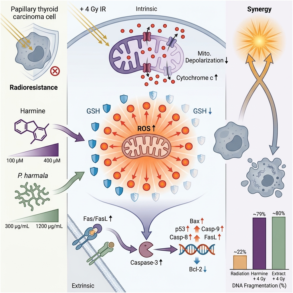
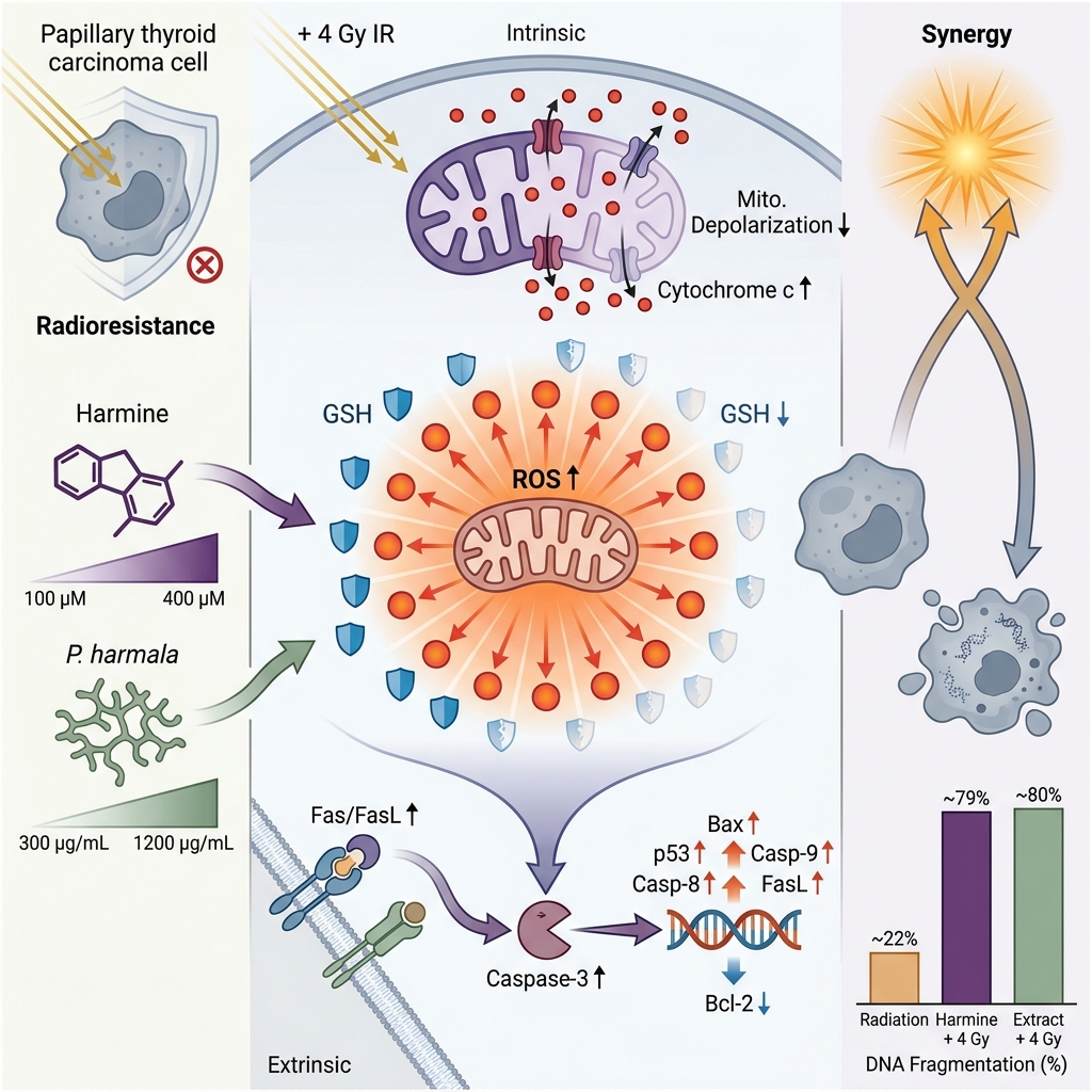
Conclusion: Harmine and P. harmala seed extract enhance radiation-induced cytotoxicity and apoptosis in B-CPAP thyroid cancer cells through oxidative stress induction, impairment of endogenous antioxidant defenses, and activation of both intrinsic (mitochondrial) and extrinsic (death-receptor) apoptotic pathways. These findings suggest that harmine and P. harmala extract represent promising natural radiosensitizing candidates that warrant further validation through clonogenic survival assays and in vivo models.
Graphical abstract
Introduction
Thyroid cancer represents the most common endocrine malignancy worldwide, and its incidence has steadily increased over recent decades.1 Although the majority of patients respond favorably to standard therapeutic interventions—including surgical resection, radioactive iodine (RAI) ablation, and thyroid-stimulating hormone (TSH) suppression therapy—radioresistant tumors remain a major clinical obstacle.2 Resistance to ionizing radiation is strongly associated with compromised treatment efficacy, persistent disease, local recurrence, and poor overall prognosis.3 Multiple biological mechanisms contribute to the radioresistant phenotype in thyroid carcinoma cells, including enhanced DNA repair capacity, dysregulated apoptotic machinery, and robust antioxidant defense networks.4 Consequently, therapeutic strategies capable of modulating or overcoming these radioresistance pathways represent an urgent clinical need.
In recent years, natural phytochemicals have received increasing attention as potential radiosensitizers due to their favorable toxicity profiles, biochemical diversity, and multi-target mechanisms of action.5Peganum harmala L. (commonly known as Syrian rue) is a perennial medicinal plant traditionally utilized in Middle Eastern and Asian medical practices. It is particularly rich in β-carboline alkaloids, most notably harmine and harmaline.6 These alkaloids possess a wide spectrum of biological activities, including antiproliferative, pro-apoptotic, anti-inflammatory, and redox-modulating effects. Harmine, in particular, has been demonstrated to inhibit tumor cell proliferation, trigger G0/G1 cell-cycle arrest, activate mitochondrial-mediated apoptotic pathways, and interfere with oncogenic signaling cascades across various malignancy models.7 High-performance liquid chromatography (HPLC) characterization has shown that P. harmala seeds contain major β-carboline alkaloids, including harmine, harmaline, harmalol, harmol, and harmane, although their relative abundance varies depending on geographical origin, harvesting conditions, and extraction procedures.8
Despite growing evidence supporting the antineoplastic properties of P. harmala constituents, their potential ability to modulate radiosensitivity in thyroid cancer has not been previously investigated. Given the pivotal role of intracellular reactive oxygen species (ROS) in mediating radiation-induced cytotoxicity, and considering the established capacity of harmine and P. harmala extracts to alter cellular redox homeostasis, these natural compounds serve as logical candidates for combination with radiotherapy.
Therefore, the present study was designed to investigate the radiosensitizing efficacy of harmine and P. harmala seed extract in human papillary thyroid cancer cells (B-CPAP). We evaluated their impact on cell viability, apoptotic death, intracellular oxidative stress markers, mitochondrial membrane integrity, and the expression of key genes governing intrinsic and extrinsic apoptotic cascades. Characterizing the molecular basis of these interactions may provide valuable insights into the utility of plant-derived radiosensitizers as adjunctive therapeutic strategies for thyroid cancer treatment.
Methods
Cell Line and Culture Conditions
The human papillary thyroid carcinoma cell line B-CPAP was obtained from the National Cell Bank of Iran (Pasteur Institute of Iran, Tehran, Iran). Cells were cultured in Dulbecco’s Modified Eagle Medium (DMEM; Gibco, Carlsbad, CA, USA) supplemented with 10% fetal bovine serum (FBS; Gibco) and 1% penicillin–streptomycin (100 U/mL penicillin, 100 µg/mL streptomycin). Cell cultures were maintained at 37 °C in a humidified incubator with a 5% CO₂ atmosphere.
Preparation of P. harmala Seed Extract
Peganum harmala seeds were finely powdered and extracted using 70% ethanol by maceration for 72 h at room temperature. The crude extract was filtered, concentrated under reduced pressure using a rotary evaporator, and dissolved in dimethyl sulfoxide (DMSO) to yield a 100 mg/mL stock solution. Working concentrations (300, 600, and 1200 µg/mL) were prepared freshly prior to each experiment by diluting the stock solution in complete culture medium. The final DMSO concentration was maintained below 0.1% (v/v) in all treatment groups, a concentration that exerted no significant effect on cell viability.9 The crude extract utilized in this study was not subjected to detailed phytochemical characterization; therefore, exact quantification of individual alkaloid constituents was not performed.
Harmine Preparation
Harmine (Sigma-Aldrich, St. Louis, MO, USA) was dissolved in DMSO to prepare a 100 mM stock solution. Experimental working concentrations (100, 200, and 400 µM) were obtained by diluting the stock in culture medium. The final vehicle (DMSO) concentration was maintained below 0.1% (v/v) across all assays.
Irradiation Procedure
Cells were exposed to a single dose of 4 Gy ionizing radiation using a calibrated linear accelerator (6 MV X-rays) at room temperature. All irradiations were conducted under standardized dosimetric conditions. Non-irradiated control cells were handled in parallel under identical ambient conditions without radiation exposure. Irradiation was administered 2 h following drug treatment to facilitate cellular uptake of the compounds.10
Cell Viability Assay and Drug–Radiation Interactions
Cell viability was evaluated using the 3-(4,5-dimethylthiazol-2-yl)-2,5-diphenyltetrazolium bromide (MTT) assay. B-CPAP cells were seeded into 96-well plates (1 × 10 cells/well) and allowed to adhere overnight. Cells were treated with specified concentrations of harmine or P. harmala extract followed by irradiation (4 Gy). After 24 h of incubation post-treatment, 20 µL of MTT solution (5 mg/mL in phosphate-buffered saline) was added to each well and incubated for 4 h at 37 °C. Formazan crystals were dissolved in 150 µL of DMSO per well, and optical density was measured at 570 nm using a microplate reader. Cell viability was expressed as a percentage relative to untreated control cells.11 Half-maximal inhibitory concentration (IC) values for harmine and P. harmala seed extract were calculated using GraphPad Prism 9 (GraphPad Software, San Diego, CA, USA). Combination Index (CI) values were determined using CompuSyn software (ComboSyn, Inc., Paramus, NJ, USA) based on the median-effect method of Chou–Talalay for multiple compound concentrations combined with a fixed 4 Gy radiation dose. Because full radiation dose–response curves were not generated, the calculated CI values represent preliminary estimates of drug–radiation interaction at the selected radiation dose rather than a complete Chou–Talalay synergy profile.
DNA Fragmentation and Apoptosis Assays
DNA fragmentation was quantified using the colorimetric diphenylamine (DPA) method. Following treatment, harvested cell pellets were lysed, and fragmented DNA was separated from intact chromatin by centrifugation. Absorbance was recorded at 600 nm, and the percentage of fragmented DNA was calculated as previously described.12
Apoptotic nuclear morphology was assessed using a Terminal deoxynucleotidyl transferase dUTP Nick End Labeling (TUNEL) detection kit (Elabscience, Wuhan, China) according to the manufacturer's instructions.13 Stained cells were visualized under a fluorescence microscope, and the apoptotic index was calculated by counting TUNEL-positive cells relative to total cells across randomly selected fields.
Measurement of Intracellular Reactive Oxygen Species
Intracellular reactive oxygen species (ROS) levels were measured using 2',7'-dichlorodihydrofluorescin diacetate (DCFH-DA; Sigma-Aldrich). Briefly, following treatment and irradiation, cells were incubated with DCFH-DA. Intracellular esterases deacetylate DCFH-DA to non-fluorescent DCFH, which is subsequently oxidized by ROS into highly fluorescent 2',7'-dichlorofluorescein (DCF). Fluorescence intensity was recorded at excitation/emission wavelengths of 485/530 nm using a microplate reader and normalized to cell number.14
Measurement of Intracellular Glutathione
Intracellular reduced glutathione (GSH) content was quantified using a commercial GSH assay kit (Sigma-Aldrich) based on the reaction with 5,5'-dithiobis(2-nitrobenzoic acid) (DTNB). Cell lysates were prepared following treatments, and absorbance was measured at 412 nm. Total GSH levels were calculated using a standard calibration curve.14
Measurement of Mitochondrial Membrane Potential
Mitochondrial membrane potential (MMP, ΔΨm) was evaluated using the cationic lipophilic dye JC-1 (Sigma-Aldrich). Following 24 h of treatment at IC concentrations, JC-1 dye (10 µM) was added and incubated for 30 min at 37 °C in the dark. Fluorescence intensity was measured at excitation/emission wavelengths of 492/520 nm (monomeric form, green) and 544/590 nm (aggregate form, red). The red-to-green (590/520 nm) fluorescence ratio was calculated to quantify relative mitochondrial membrane potential.14
Measurement of Cytochrome c Release
Cytosolic cytochrome c levels were quantified using a human Cytochrome c ELISA Kit (Abcam, Cambridge, MA, USA) according to the manufacturer’s protocol. Absorbance was recorded at 450 nm using a microplate reader.14
Gene Expression Analysis
Total RNA was extracted from cells using an RNA isolation kit (Life Technologies, Carlsbad, CA, USA). Complementary DNA (cDNA) was synthesized from 1 µg of total RNA using a reverse transcription kit (Takara Bio, Otsu, Japan). Quantitative real-time PCR (qRT-PCR) was performed with SYBR Green Master Mix (Applied Biosystems, Foster City, CA, USA) on a StepOnePlus™ Real-Time PCR System. Thermal cycling conditions comprised an initial step at 50 °C for 10 min, hot-start activation at 95 °C for 10 min, followed by 40 cycles of denaturation at 95 °C for 15 s and annealing/extension at 60 °C for 45 s. Specific primer sequences for Bax, Bcl-2, p53, Caspase-3, Caspase-8, Caspase-9, Fas, FasL, and GAPDH are listed in Table 1. Relative mRNA expression levels were calculated using the 2 comparative threshold method, with GAPDH serving as the endogenous reference gene.14
Primer sequences used for quantitative real-time PCR (qRT-PCR) analysis.
| Gene | Forward Primer (5'–3') | Reverse Primer (5'–3') |
|---|---|---|
| TTCTGCCATAAGCCCTGTCC | TGTACTCCTTCCCTTCTTGG | |
| GCCTGTGTCTCCTTGTGATG | TGGACTTGCCTGTTAAATGGG | |
| AGTAACATGGAGCTGCAGAGGAT | GCTGCCACTCGGAAAAAGAC | |
| TGGCCAGGGTCAGAGTTAAA | TGGCCTCTCTTGCGGAGTA | |
| CCTCAGCATCTTATCCGAGTGG | TGGATGGTGGTACAGTCAGAGC | |
| GGAAGCGAATCAATGGACTCTGG | GCATCGACATCTGTACCAGACC | |
| AGAAGAGGGTCATCCTGGGAGA | TCAGGACTTCCTTCAAGGCTGC | |
| GTTTGAGGACCTTCGACCAGCT | CAACGTACCAGGAGCCACTCTT | |
| TGTGGGCATCAATGGATTTGG | ACACCATGTATTCCGGGTCAAT |
Statistical Analysis
All experiments were performed independently in triplicate (biological replicates), with each sample analyzed in technical triplicate. Data are presented as mean ± standard deviation (SD). Statistical analyses were conducted using one-way Analysis of Variance (ANOVA) followed by Tukey’s post-hoc test for multiple comparisons in GraphPad Prism 9. Differences were considered statistically significant at p < 0.05.
Results
Effect of Harmine and P. harmala Extract on Cell Viability
Treatment of B-CPAP thyroid cancer cells with increasing concentrations of harmine (100, 200, and 400 µM) or P. harmala seed extract (300, 600, and 1200 µg/mL) resulted in a significant, dose-dependent decrease in cell viability. The calculated IC values were 1053.57 ± 22.96 µg/mL for P. harmala seed extract and 328.12 ± 11.66 µM for harmine. Exposure to 4 Gy ionizing radiation alone produced moderate cytotoxicity (83.00 ± 2.89% viability). However, combination treatment of radiation with either harmine or P. harmala extract significantly enhanced growth inhibition compared with single-agent or radiation-alone groups (p < 0.05). The most substantial reduction in viability occurred in cells co-treated with 400 µM harmine + 4 Gy radiation (24.00 ± 1.00% viability) and 1200 µg/mL extract + 4 Gy radiation (20.00 ± 2.00% viability; Figure 1A and 1B).
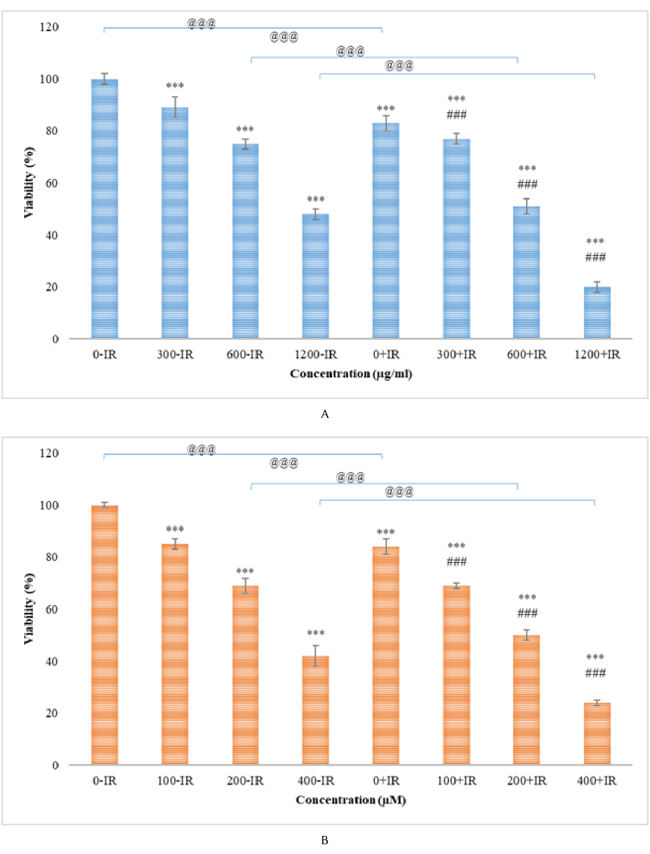
Effects of harmine and
The doses of harmine, P. harmala extract, and ionizing radiation used in combination treatments, along with the corresponding CI values calculated using the Chou–Talalay method, are summarized in Table 2. For harmine at doses of 100, 200, and 400 µM combined with 4 Gy radiation, the calculated CI values were 0.93, 0.77, and 0.60, respectively (Table 2), indicating moderate to strong synergistic interactions at higher concentrations. For P. harmala seed extract at doses of 300, 600, and 1200 µg/mL combined with 4 Gy radiation, CI values were 1.21, 0.79, and 0.26, respectively (Table 2). These preliminary CI values demonstrate potential synergistic interactions between the tested natural compounds and ionizing radiation at the selected dose.
Combination Index (CI) values for harmine and
| Treatment Agent | Combination Concentration / Dose | Combination Index (CI)* | Interaction Description |
|---|---|---|---|
| Harmine | 100 µM + 4 Gy | 0.93 | Slight synergy |
| 200 µM + 4 Gy | 0.77 | Moderate synergy | |
| 400 µM + 4 Gy | 0.60 | Synergy | |
| 300 µg/mL + 4 Gy | 1.21 | Slight antagonism | |
| 600 µg/mL + 4 Gy | 0.79 | Moderate synergy | |
| 1200 µg/mL + 4 Gy | 0.26 | Strong synergy |
Harmine and P. harmala Extract Potentiate Radiation-Induced Apoptosis
Both harmine and P. harmala extract increased DNA fragmentation in a concentration-dependent manner. Radiation alone moderately elevated DNA fragmentation (22.33 ± 4.16%), whereas co-treatment with P. harmala extract + 4 Gy (75.33 ± 2.52%) or harmine + 4 Gy (79.33 ± 3.21%) markedly amplified apoptotic DNA fragmentation compared with all other groups (p < 0.05; Figure 2B). Microscopic evaluation of TUNEL-stained cells demonstrated a significant increase in TUNEL-positive nuclei following treatment with either agent. Combination therapy with radiation yielded the highest apoptotic index, confirming synergistic apoptosis induction (Figure 2A).
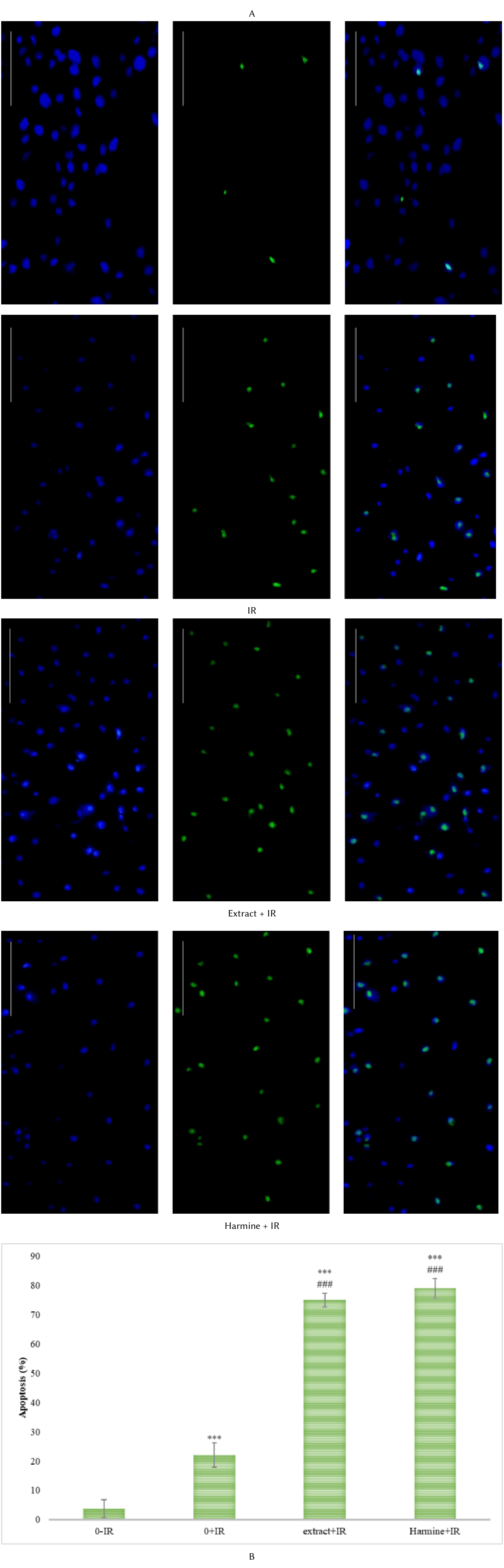
Effects of harmine and
Effects on Intracellular Reactive Oxygen Species Levels
Harmine and P. harmala extract significantly elevated intracellular ROS generation compared with untreated controls (p < 0.05). Exposure to 4 Gy radiation alone also increased ROS production (1.94-fold relative to control). However, combined treatment induced a dramatic surge in ROS levels (2.67-fold for extract + 4 Gy; 3.00-fold for harmine + 4 Gy), which was significantly higher than either individual treatment (p < 0.05). The highest ROS accumulation occurred in the high-dose harmine + radiation group, indicating that heightened oxidative stress plays a critical role in their radiosensitizing mechanism (Figure 3A).
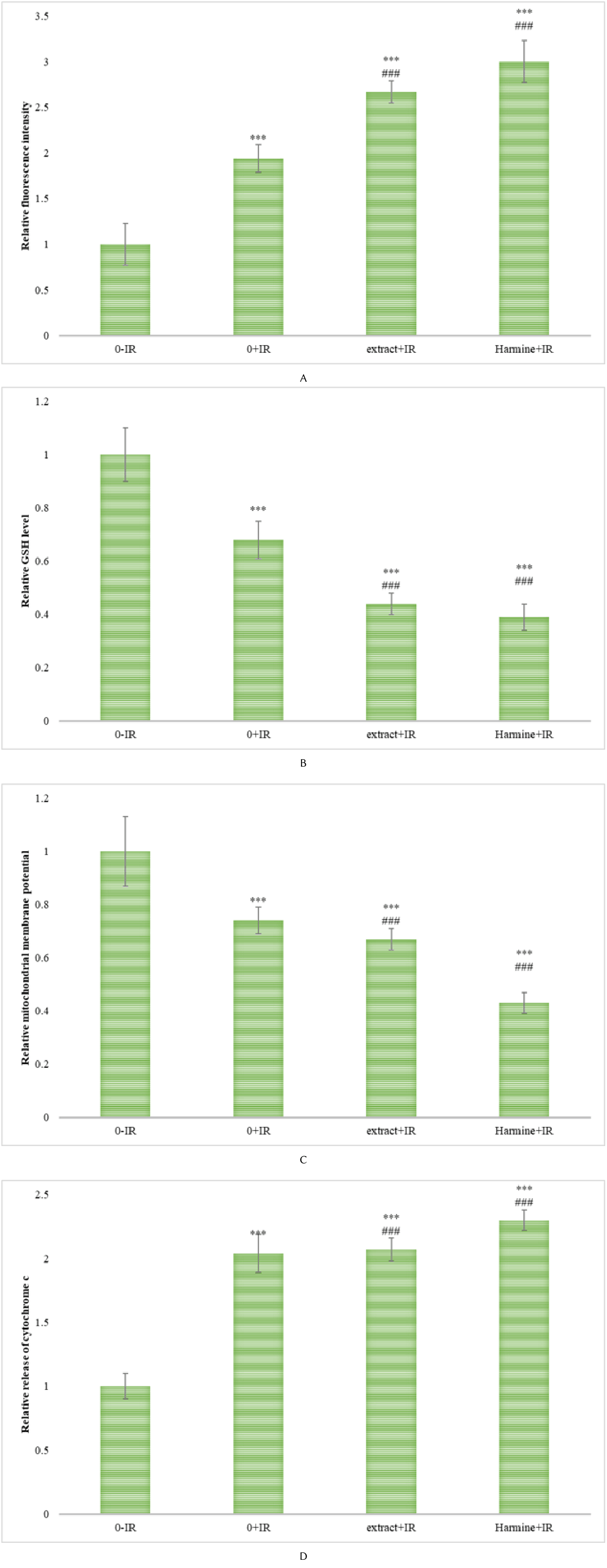
Effects of harmine and
Effects on Cellular Glutathione Levels
Both natural agents induced a dose-dependent depletion of intracellular GSH, indicating compromised cellular antioxidant capacity. Radiation alone moderately reduced GSH levels (0.68-fold of control). Co-treatment with harmine or P. harmala extract and radiation caused the most severe GSH depletion (0.44-fold for extract + 4 Gy; 0.39-fold for harmine + 4 Gy; p < 0.05), inversely correlating with ROS elevation (Figure 3B).
Effect on Mitochondrial Membrane Potential
Mitochondrial membrane potential (MMP) was significantly reduced in cells treated with harmine or P. harmala seed extract (p < 0.05). This depolarization was significantly amplified upon co-treatment with 4 Gy ionizing radiation (0.67-fold for extract + 4 Gy; 0.43-fold for harmine + 4 Gy relative to control; p < 0.05; Figure 3C).
Effect on Cytochrome c Release
Cytosolic cytochrome c levels were significantly increased following harmine or P. harmala extract exposure (p < 0.05). The most prominent accumulation of cytosolic cytochrome c was observed in the radiation co-treatment groups (2.07-fold for extract + 4 Gy; 2.30-fold for harmine + 4 Gy relative to control; p < 0.05; Figure 3D).
Modulation of Apoptosis-Related Gene Expression
qRT-PCR analysis revealed that combined treatment with harmine or P. harmala extract and radiation significantly upregulated pro-apoptotic genes, including Bax, p53, Caspase-3, Caspase-8, Caspase-9, Fas, and FasL (p < 0.05; Figure 4A). Expression levels peaked in groups receiving the highest compound doses in combination with 4 Gy radiation. Conversely, expression of the anti-apoptotic gene Bcl-2 was significantly suppressed across all treated groups, with the most profound downregulation observed in co-treated cells (p < 0.05; Figure 4B). This expression pattern strongly supports the concurrent activation of intrinsic (mitochondrial) and extrinsic (death-receptor) apoptotic pathways.
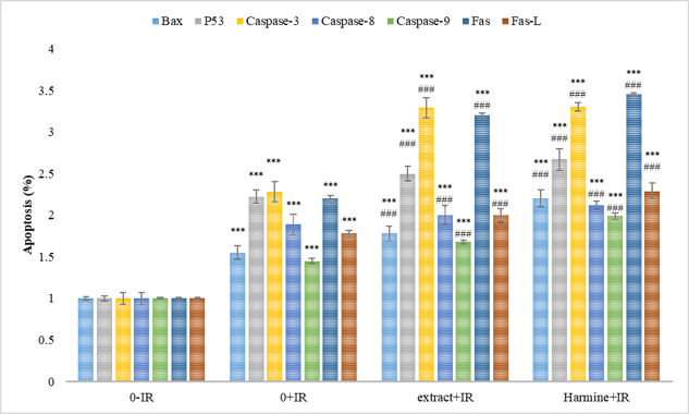
Modulation of apoptosis-related gene expression by harmine and
Discussion
The present study demonstrates that harmine and P. harmala seed extract exert potent cytotoxic and pro-apoptotic activities in B-CPAP thyroid cancer cells. Crucially, combining these natural compounds with ionizing radiation significantly enhanced radiation-induced cytotoxicity, demonstrating a synergistic radiosensitizing effect. Combination Index analysis confirmed that harmine and P. harmala extract exhibit synergistic interactions with 4 Gy radiation at specific concentrations, providing quantitative evidence of enhanced efficacy at the cellular level.
Radioresistance represents a primary therapeutic barrier in thyroid oncology and is typically driven by augmented cell survival pathways, robust DNA repair mechanisms, evasion of apoptosis, and efficient redox buffering capacity.15 Radiosensitizers act by disabling these cytoprotective adaptations, thereby rendering tumor cells heightened sensitivity to radiation damage.16 The dose-dependent reduction in cell viability observed with harmine or P. harmala extract alone aligns with previous studies documenting the antiproliferative and pro-apoptotic activities of β-carboline alkaloids in various cancer models.17 However, the marked potentiated cytotoxicity when combined with ionizing radiation reflects true synergistic interaction rather than simple additive effect.
The pronounced growth inhibition observed with 400 µM harmine or 1200 µg/mL P. harmala extract in combination with radiation suggests that achieving threshold intracellular drug accumulation is essential for optimal radiosensitization. This finding is consistent with earlier reports demonstrating that natural phytochemicals can sensitize malignant cells to radiation in a dose-dependent manner by disrupting radiation-induced stress responses.18 Importantly, while these findings establish radiosensitization in B-CPAP thyroid cancer cells, non-tumor thyroid epithelial cells were not examined in this study; thus, definitive conclusions regarding therapeutic selectivity and normal tissue toxicity cannot be drawn.
Our results show that harmine and P. harmala extract significantly induce apoptosis in B-CPAP cells, as confirmed by elevated DNA fragmentation and an increased percentage of TUNEL-positive nuclei, whereas radiation alone produced only moderate apoptotic death. Combination of radiation with harmine or P. harmala extract markedly amplified apoptotic responses.
DNA fragmentation is a classic biochemical hallmark of late-stage apoptosis, reflecting irreversible commitment to programmed cell death.19 The substantial elevation in fragmented DNA following co-treatment indicates that harmine and P. harmala extract actively augment radiation-induced apoptotic signaling cascades rather than inducing non-specific necrosis. This conclusion is reinforced by TUNEL assay results showing pronounced accumulation of apoptotic nuclei. These findings accord with prior research demonstrating that harmine can trigger apoptotic cascades via mitochondrial disruption and caspase activation in diverse neoplastic models.20,21,22,23,24,25,26,27
The synergistic induction of apoptosis observed here may stem from the ability of harmine and P. harmala-derived alkaloids to disrupt cellular defense mechanisms that normally limit radiation-induced cell death. Radioresistance in thyroid cancer has been repeatedly linked to impaired apoptotic execution and upregulation of pro-survival signaling.28 By lowering the threshold for apoptotic activation, harmine and P. harmala extract effectively bypass these resistance mechanisms, thereby enhancing radiation sensitivity.
Oxidative stress is a primary driver of radiation-induced cell death, occurring through excessive ROS production that causes damaging lesions in cellular macromolecules, including DNA, proteins, and membrane lipids.29 In this study, harmine and P. harmala extract significantly elevated intracellular ROS levels, an effect dramatically intensified upon co-treatment with 4 Gy radiation. This marked ROS accumulation in co-treated cells indicates that oxidative stress serves as a central mediator of their radiosensitizing properties.
Radiation-induced ROS generation is normally counteracted by endogenous antioxidant systems, among which intracellular GSH serves as the principal non-enzymatic scavenger.30 We found that harmine and P. harmala extract significantly depleted intracellular GSH pools, particularly when combined with radiation. The simultaneous elevation of ROS and depletion of GSH strongly suggest that these agents collapse cellular redox homeostasis, rendering cells incapable of detoxifying radiation-induced free radicals.
This exacerbated oxidative stress directly fuels cytotoxicity and apoptosis. High ROS levels induce mitochondrial membrane permeabilization, exacerbate DNA double-strand breaks, and activate redox-sensitive pro-apoptotic signaling pathways. Simultaneously, GSH depletion impairs radical scavenging, creating a highly permissive redox environment for apoptotic execution. Previous studies have noted that harmine modulates oxidative stress by influencing mitochondrial function and redox-sensitive enzymes,31 while P. harmala alkaloids impair antioxidant defenses in cancer cells.32 Our findings extend these observations by demonstrating that harmine and P. harmala extract potentate radiation-induced oxidative damage in thyroid cancer cells.
Mitochondrial dysfunction represents a crucial event in radiation-induced apoptosis and a primary determinant of cellular radiosensitivity.33 Here, treatment with harmine or P. harmala extract significantly reduced mitochondrial membrane potential (MMP), an effect further exacerbated upon co-treatment with radiation. Loss of MMP is a recognized indicator of outer mitochondrial membrane permeabilization, representing an irreversible early commitment step in intrinsic apoptosis.34 The pronounced depolarization in co-treated cells confirms that harmine and P. harmala extract disrupt mitochondrial integrity, thereby amplifying radiation-induced apoptotic cascades.
Consistent with mitochondrial depolarization, cytosolic cytochrome c levels were significantly elevated in harmine- and P. harmala-treated cells, reaching peak levels in combination with radiation. Cytochrome c release into the cytosol triggers apoptosome assembly and activates downstream executioner caspases.35 These data provide concrete mechanistic evidence that mitochondrial-mediated intrinsic apoptosis drives the observed radiosensitization.
At the transcriptional level, gene expression analysis further confirmed the engagement of both intrinsic and extrinsic apoptotic pathways. Combined treatment significantly upregulated pro-apoptotic genes (Bax, p53, Caspase-3, Caspase-8, Caspase-9, Fas, and FasL) while simultaneously suppressing anti-apoptotic Bcl-2 expression. The resulting elevation in the Bax/Bcl-2 ratio favors mitochondrial outer membrane permeabilization, shifting the cellular balance toward death. Concurrent upregulation of Caspase-9 and Caspase-3 confirms execution of the intrinsic pathway, whereas elevated expression of Caspase-8, Fas, and FasL demonstrates activation of the death receptor-mediated extrinsic pathway. This dual activation highlights the capacity of harmine and P. harmala extract to recruit multiple apoptotic pathways, overcoming single-pathway resistance mechanisms.
Several study limitations should be acknowledged. First, investigations were conducted on a single papillary thyroid cancer cell line (B-CPAP) without parallel evaluation in normal thyroid epithelial cells. Second, the P. harmala seed extract was crude and not chemically profiled using HPLC or LC-MS/MS. Third, irradiation was evaluated at a single dose (4 Gy), clonogenic survival assays were not performed, and CI calculations were restricted to a single radiation dose. Therefore, formal validation of radiosensitization, phytochemical standardization, detailed synergy mapping, and in vivo translational efficacy remain essential goals for future research.
Conclusion
In summary, harmine and P. harmala seed extract markedly enhance radiation-induced cytotoxicity and apoptosis in B-CPAP papillary thyroid cancer cells through a multi-target mechanism involving oxidative stress induction, GSH depletion, mitochondrial membrane depolarization, cytosolic cytochrome c release, and concurrent activation of intrinsic and extrinsic apoptotic signaling cascades. These natural agents represent promising candidate radiosensitizers for thyroid cancer management, warranting further evaluation through clonogenic survival assays, chemical standardization, and in vivo preclinical studies.
Declarations
Abbreviations
ANOVA: Analysis of Variance; Bax: Bcl-2-associated X protein; Bcl-2: B-cell lymphoma 2; B-CPAP: Human papillary thyroid carcinoma cell line; cDNA: Complementary DNA; CI: Combination Index; DCFH-DA: 2',7'-Dichlorodihydrofluorescin diacetate; DMEM: Dulbecco’s Modified Eagle Medium; DMSO: Dimethyl sulfoxide; DPA: Diphenylamine; DTNB: 5,5'-Dithiobis(2-nitrobenzoic acid); ELISA: Enzyme-linked immunosorbent assay; Fas: Fas cell surface death receptor; FasL: Fas ligand; FBS: Fetal bovine serum; GAPDH: Glyceraldehyde 3-phosphate dehydrogenase; GSH: Reduced glutathione; Gy: Gray (unit of absorbed radiation dose); HPLC: High-performance liquid chromatography; IC: Half-maximal inhibitory concentration; JC-1: 5,5',6,6'-Tetrachloro-1,1',3,3'-tetraethylbenzimidazolylcarbocyanine iodide; LC-MS/MS: Liquid chromatography-tandem mass spectrometry; MMP: Mitochondrial membrane potential; MTT: 3-(4,5-Dimethylthiazol-2-yl)-2,5-diphenyltetrazolium bromide; PCR: Polymerase chain reaction; qRT-PCR: Quantitative real-time polymerase chain reaction; ROS: Reactive oxygen species; SD: Standard deviation; TSH: Thyroid-stimulating hormone; TUNEL: Terminal deoxynucleotidyl transferase dUTP nick end labeling.
Acknowledgments
The authors sincerely thank Kermanshah University of Medical Sciences for providing laboratory facilities, equipment, and technical support throughout this research project.
Author’s contributions
RN: Conceptualization, Formal analysis, Statistical analysis, Writing – original draft. CJ: Project administration, Data curation, Supervision, Writing – original draft, Writing – review & editing. MP: Cell culture, Laboratory experiments, Methodology. RK: Validation, Investigation, Writing – review & editing. All authors read and approved the final manuscript.
Funding
This study was financially supported by the Vice-Chancellor for Research at Kermanshah University of Medical Sciences (Project Grant No. 4030393).
Availability of data and materials
The datasets generated and/or analyzed during the current study are available from the corresponding author upon reasonable request.
Ethics approval and consent to participate
Ethics approval: The study protocol was approved by the Ethics Committee of Kermanshah University of Medical Sciences, Kermanshah, Iran (Approval Code: IR.KUMS.MED.REC.1403.141).
Consent to participate: Not applicable (this study was conducted exclusively on an in vitro cell line and did not involve human participants or animal tissue).
Consent for publication
Not applicable.
Declaration of generative AI and AI-assisted technologies in the writing process
During the preparation of this manuscript, the authors used an AI-assisted writing tool to improve the language, grammar, and readability of the text. The AI tool was used solely for writing assistance and did not contribute to the scientific content, study design, data analysis, interpretation of results, or conclusions. The authors carefully reviewed and edited the final manuscript and take full responsibility for its content.
Competing interests
The authors declare that they have no competing interests.