


Macroalga Sargassum polycystum-Mediated Antiproliferation and Apoptosis in MCF-7 Breast Cancer Cell Lines

- PG & Research Department of Biochemistry, Mohamed Sathak College of Arts and Science, Greater Chennai, Tamil Nadu 600119, India
- PG & Research Department of Biotechnology, Mohamed Sathak College of Arts and Science, Greater Chennai, Tamil Nadu 600119, India
Abstract
Background & Objective: Marine brown algae are rich natural sources of bioactive metabolites with substantial therapeutic potential. Sargassum polycystum, abundantly distributed along the Indian coastline, has received limited systematic evaluation regarding its pharmacological properties. The objective of the present study was to comprehensively investigate the phytochemical profile, pigment composition, antioxidant, anti-inflammatory, and anticancer activities of S. polycystum.
Methods: Ethanolic and hexane extracts were prepared and subjected to qualitative phytochemical screening. The predominant chemical constituents in the ethanolic extract were characterized using gas chromatography–mass spectrometry (GC–MS) and high-performance liquid chromatography (HPLC), alongside quantification of total phenolics and flavonoids. Photosynthetic pigments extracted using ethanol, acetone, chloroform, and ethyl acetate were separated by thin-layer chromatography (TLC), and pigment concentrations in ethanolic and ethyl acetate extracts were quantified spectrophotometrically. Antioxidant potential was evaluated using total antioxidant capacity (TAC), 2,2-diphenyl-1-picrylhydrazyl (DPPH), and 2,2'-azino-bis(3-ethylbenzothiazoline-6-sulfonic acid) (ABTS) radical scavenging assays. Anti-inflammatory activity was assessed via human red blood cell (HRBC) membrane stabilization. Anticancer potential was evaluated in vitro against MCF-7 breast cancer cell lines through the 3-(4,5-dimethylthiazol-2-yl)-2,5-diphenyltetrazolium bromide (MTT) cytotoxicity assay, genomic DNA fragmentation analysis, and reverse transcription-polymerase chain reaction (RT-PCR) expression analysis of the p53 tumor suppressor gene.
Results: Phytochemical screening revealed diverse secondary metabolites, and pigment profiles were confirmed by TLC, GC–MS, and HPLC chromatograms. The ethanolic extract possessed high concentrations of total phenolics (2.13 ± 0.36 mg GAE g⁻¹ dw) and flavonoids (2.49 ± 0.42 mg RE g⁻¹ dw). The extract demonstrated potent, dose-dependent antioxidant and anti-inflammatory activities comparable to standard reference drugs. Furthermore, S. polycystum ethanolic extract suppressed MCF-7 cancer cell viability (IC₅₀ = 104.3 µg/mL), induced characteristic internucleosomal DNA laddering, and markedly upregulated p53 mRNA expression.
Conclusion: These findings demonstrate that S. polycystum is a promising marine resource containing diverse bioactive constituents capable of inducing p53-mediated apoptosis in breast cancer cells, highlighting its potential for the development of novel natural therapeutic agents.
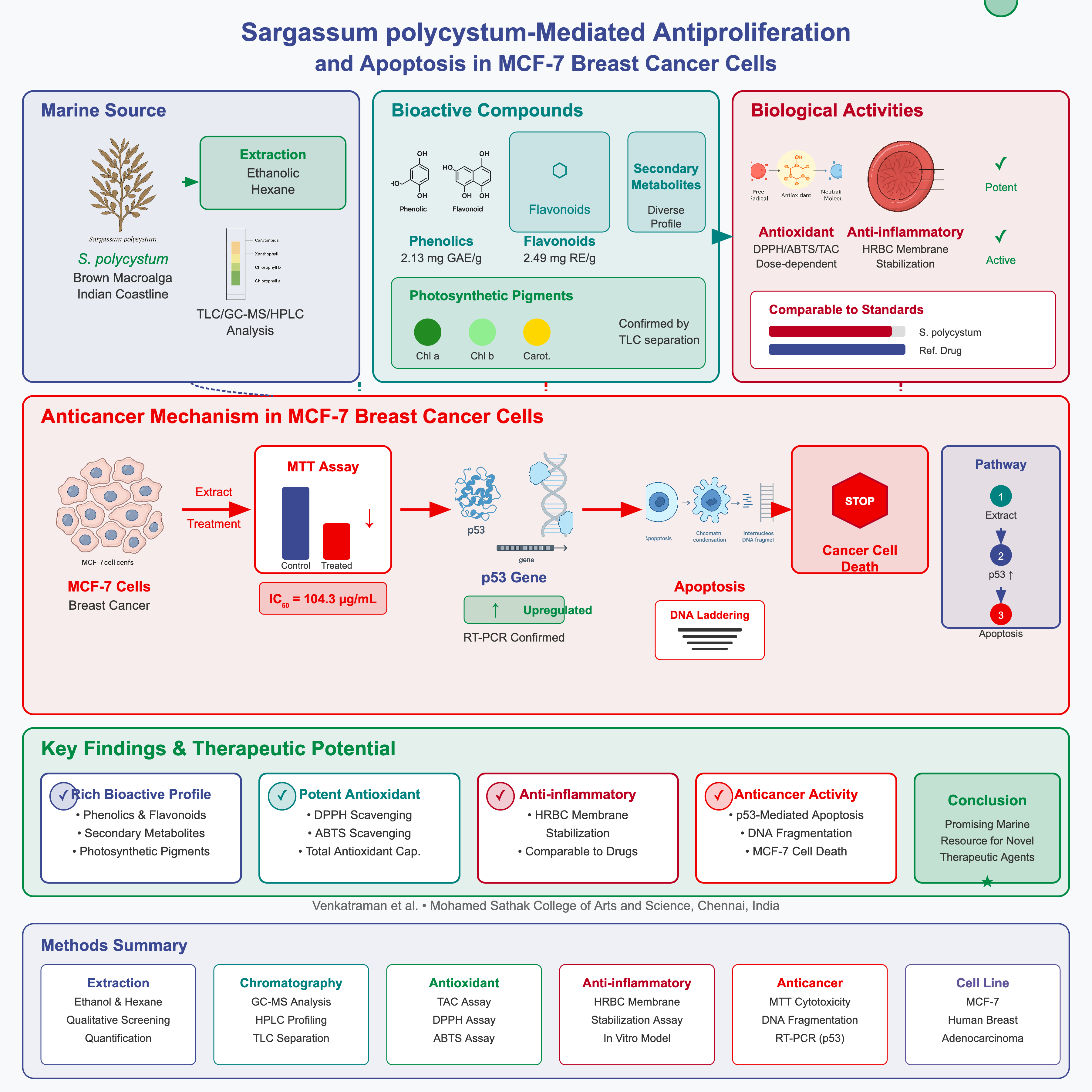
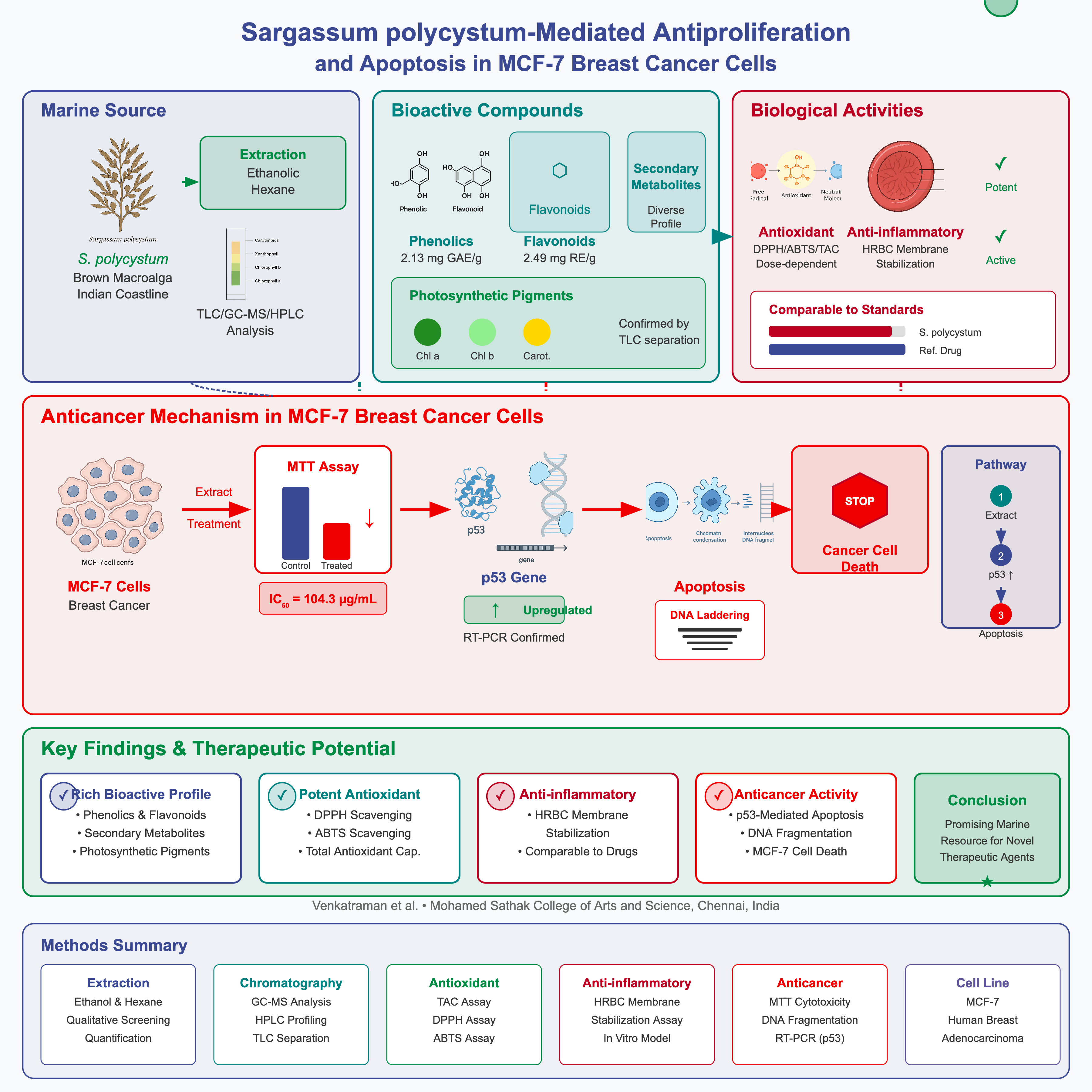
Graphical abstract
INTRODUCTION
Marine macroalgae are increasingly recognized as valuable natural resources yielding structurally diverse bioactive compounds. Among them, brown seaweeds (Phaeophyceae) are particularly rich in polysaccharides, polyphenols, pigments, fatty acids, and secondary metabolites that possess potent antioxidant, antimicrobial, anti-inflammatory, and cytotoxic properties1,2. Consequently, marine macroalgae find diverse applications in pharmaceuticals, nutraceuticals, and functional foods.
The genus Sargassum is among the most abundant brown algae distributed throughout tropical and subtropical marine ecosystems. Species of Sargassum have been reported to exhibit a wide array of biological activities, including free-radical scavenging, immunomodulatory, hypoglycemic, antimicrobial, analgesic, anti-inflammatory, and anticancer effects. These properties are largely attributed to phytoconstituents such as fucoxanthin, phlorotannins, sterols, and sulfated polysaccharides3,4,5,6,7.
Cancer remains one of the leading causes of mortality worldwide. Conventional chemotherapeutic agents are frequently limited by systemic toxicity, severe side effects, and drug resistance arising during prolonged treatment8. Natural products derived from marine organisms continue to play a pivotal role in anticancer drug discovery by providing novel chemical scaffolds capable of selectively inducing apoptosis in malignant cells with reduced adverse effects9,10. Algae-derived compounds have demonstrated promising anticancer mechanisms, including cell-cycle arrest, mitochondrial dysfunction, DNA fragmentation, and modulation of key tumor suppressor genes such as p5311,12.
In addition to tumor growth and metastasis, oxidative stress and cellular inflammation are closely linked to the pathogenesis and progression of chronic human diseases, including cancer, cardiovascular disorders, and neurodegenerative conditions. Antioxidants from marine algae neutralize reactive oxygen species (ROS) and prevent cellular oxidative damage, whereas anti-inflammatory constituents stabilize cell membranes and inhibit heat-induced protein denaturation13,14.
Although Sargassum polycystum is widely distributed along the Gulf of Mannar, systematic investigations integrating detailed phytochemical characterization with comprehensive antioxidant, anti-inflammatory, and anticancer evaluations remain limited. Therefore, the present study aimed to comprehensively evaluate the bioactive potential of S. polycystum using chromatographic characterization and in vitro bioassays.
MATERIALS AND METHODS
Collection and Authentication of Algal Material
Healthy specimens of Sargassum polycystum were collected from the coastal region of Mandapam, Gulf of Mannar, Rameswaram, Tamil Nadu, India. The collected samples were washed thoroughly with seawater followed by tap water and distilled water to remove epiphytes, sand, and extraneous debris. The seaweed was shade-dried at room temperature and ground into a fine powder. Taxonomic identification was confirmed using standard seaweed manuals, and authentication was performed by marine algal taxonomist Dr. Murali, Professor, Department of Plant Biology and Biotechnology, Government Arts College, Nandanam, Chennai, Tamil Nadu, India15.
Preparation of Extracts
Ten grams of shade-dried powdered algal material was extracted separately with ethanol and hexane (1:10, w/v). Each mixture was maintained in a water bath at 40°C with continuous agitation for 24 h to facilitate the extraction of phytoconstituents. The mixtures were cooled to room temperature and filtered through Whatman No. 1 filter paper. The filtrates were concentrated under reduced pressure using a rotary evaporator and stored at 4°C until further analysis16.
Phytochemical Screening
Qualitative phytochemical screening of the ethanolic and hexane extracts of S. polycystum was performed to detect alkaloids, phenols, tannins, flavonoids, steroids, terpenoids, saponins, proteins, glycosides, and carbohydrates following standard analytical protocols described by Harborne (1973)17. Although both ethanolic and hexane extracts were screened, the ethanolic extract was selected for detailed chromatographic characterization and biological evaluation due to its higher extraction yield, richer phenolic and pigment content, and superior preliminary bioactivity.
GC–MS and HPLC Analysis
Gas chromatography–mass spectrometry (GC–MS) analysis of the ethanolic extract of S. polycystum was carried out using an Agilent 5890 series II GC coupled with an Agilent 5975C inert mass selective detector equipped with an HP-5MS capillary column (30 m × 0.25 mm ID × 0.25 µm film thickness). Helium was used as the carrier gas at a constant flow rate of 1.0 mL min. The injector temperature was set at 280°C with a split ratio of 1:25. The oven temperature program was initiated at 60°C (held for 2 min), increased to 260°C at a rate of 10°C min, and held at 260°C for 10 min. Mass spectra were acquired in positive electron ionization (EI) mode at 70 eV over an m/z scan range of 40–550. Chemical constituents were identified by comparing their mass spectra with reference entries in the Wiley mass spectral library database.
High-performance liquid chromatography (HPLC) analysis was performed on a Shimadzu LC-20AD system equipped with a UV/Vis detector and a C18 reverse-phase column. Isocratic elution was conducted at a flow rate of 1.0 mL min, and major pigment compounds were identified based on retention times and spectral characteristics.
Estimation of Total Phenolics and Flavonoids
Total phenolic content (TPC) was determined using the Folin–Ciocalteu colorimetric method with gallic acid as the reference standard and expressed as mg gallic acid equivalents per gram of dry weight (mg GAE g dw)22. Total flavonoid content (TFC) was estimated using the aluminum chloride colorimetric assay with rutin as the reference standard and expressed as mg rutin equivalents per gram of dry weight (mg RE g dw)23. All assays were conducted in triplicate.
Analysis of Pigment Composition
Thin-layer chromatography (TLC) was performed to separate and profile the photosynthetic pigments of S. polycystum. Pigments were extracted individually using ethanol, acetone, chloroform, and ethyl acetate to evaluate solvent solubility. Pre-coated silica gel 60 F plates (0.25 mm thickness; Merck) were activated at 110°C for 30 min prior to use. Crude extracts (10 mg mL) were applied as 2–5 µL spots 1 cm from the base of the plate. Plates were developed in a pre-saturated chamber using a mobile phase of hexane:diethyl ether:acetone (6:3:2, v/v/v). Separated pigment bands were visualized under visible and ultraviolet (UV) light. Retention factor (Rf) values were calculated and compared with reported pigment standards18.
Photosynthetic pigments extracted from powdered S. polycystum using ethanol and ethyl acetate were quantified spectrophotometrically. Chlorophyll a, chlorophyll c + c, total chlorophyll, total carotenoids, and fucoxanthin concentrations were calculated using established spectrophotometric equations based on specific absorption wavelengths19,20,21.
In Vitro Biological Assays
Based on its higher extraction yield and rich content of polar bioactive metabolites, the ethanolic extract of S. polycystum was utilized for all subsequent in vitro antioxidant, anti-inflammatory, and anticancer assays.
Antioxidant Activity Assays
The antioxidant potential of the ethanolic extract was evaluated using three complementary in vitro methods: total antioxidant capacity (TAC), DPPH radical scavenging, and ABTS radical cation decolorization assays.
Total antioxidant capacity was determined by the phosphomolybdenum method, based on the reduction of Mo(VI) to Mo(V) by the extract and subsequent formation of a green phosphate/Mo(V) complex, with absorbance measured spectrophotometrically at 695 nm24. Ascorbic acid served as the reference standard.
DPPH (2,2-diphenyl-1-picrylhydrazyl) radical scavenging activity was assessed by monitoring the decrease in absorbance at 517 nm following reaction with various concentrations of the extract (100–500 µg/mL)25. The percentage of DPPH scavenging and half-maximal inhibitory concentration (IC) values were calculated.
The ABTS assay was carried out using pre-formed ABTS•⁺ radical cations generated by reacting 7 mM ABTS solution with 2.45 mM potassium persulfate. The reduction in absorbance was measured at 745 nm after incubation with the extract26. Ascorbic acid was used as the reference standard in all antioxidant assays, and results were expressed as mean percentage inhibition and IC values from triplicate experiments.
In Vitro Anti-Inflammatory Activity
The human red blood cell (HRBC) membrane stabilization method was employed to evaluate in vitro anti-inflammatory activity27,28. Blood was collected from a healthy human volunteer who had not taken any non-steroidal anti-inflammatory drugs (NSAIDs) for at least two weeks prior to the experiment. The blood sample was mixed with an equal volume of sterile Alsever solution (2% dextrose, 0.8% sodium citrate, 0.5% citric acid, and 0.42% NaCl), centrifuged at 3,000 rpm, and the packed red blood cells were washed three times with isotonic saline (0.9% NaCl, pH 7.2) to prepare a 10% (v/v) HRBC suspension.
The reaction mixture contained 0.5 mL of test sample (100–500 µg/mL), 1.0 mL of phosphate buffer (0.15 M, pH 7.4), 2.0 mL of hypotonic saline (0.36% NaCl), and 0.5 mL of 10% HRBC suspension. The mixture was incubated at 37°C for 30 min and centrifuged at 3,000 rpm for 20 min. The absorbance of the supernatant was measured spectrophotometrically at 560 nm. The control contained distilled water instead of the test extract. Diclofenac sodium (100–500 µg/mL) served as the standard reference drug. The percentage of membrane protection was calculated using the formula:
Cell Culture and MTT Cytotoxicity Assay
MCF-7 human breast adenocarcinoma cells (ATCC® HTB-22™) were cultured in Dulbecco’s Modified Eagle Medium (DMEM) supplemented with 10% fetal bovine serum (FBS), 100 U/mL penicillin, and 100 µg/mL streptomycin. Cells were maintained at 37°C in a humidified incubator containing 5% CO and utilized between passages 5 and 20.
Antiproliferative activity was evaluated using the 3-(4,5-dimethylthiazol-2-yl)-2,5-diphenyltetrazolium bromide (MTT) assay29. MCF-7 cells were seeded in 96-well plates at a density of 1 × 10⁵ cells/well and allowed to adhere overnight. Confluent cells were treated with various concentrations of S. polycystum ethanolic extract (SAR; 25, 50, 100, 200, and 320 µg/mL) for 24 h. Untreated cells served as the negative control. Following incubation, 20 µL of MTT solution (5 mg/mL in PBS) was added to each well and incubated at 37°C for 4 h. The resulting purple formazan crystals were dissolved in 50 µL of dimethyl sulfoxide (DMSO) with gentle agitation for 10 min. Absorbance was recorded at 570 nm using a microplate reader. Percentage cell viability was calculated as follows:
Morphological alterations in treated and control cells were documented under an inverted phase-contrast light microscope. IC values were determined by non-linear regression analysis.
DNA Fragmentation Assay
To determine whether cytotoxic cell death occurred via apoptosis, genomic DNA cleavage was analyzed by agarose gel electrophoresis11. Internucleosomal cleavage of genomic DNA into ~180–200 bp fragments is a biochemical hallmark of apoptosis. MCF-7 cells treated with different concentrations of SAR (0, 25, 50, and 100 µg/mL) or Doxorubicin (positive control) for 24 h were harvested by trypsinization and washed with phosphate-buffered saline (PBS).
Cell pellets were lysed in 500 µL of ice-cold lysis buffer containing 10 mM Tris-HCl (pH 7.4), 150 mM NaCl, 5 mM EDTA, and 0.5% Triton X-100 for 30 min on ice. Lysates were vortexed and cleared by centrifugation at 10,000 × g for 20 min at 4°C. Fragmented DNA in the supernatant was extracted using a neutral phenol:chloroform:isoamyl alcohol mixture (25:24:1, v/v/v). Purified DNA samples mixed with 6X gel loading dye were resolved on a 1.5% agarose gel containing 0.1 µg/mL ethidium bromide alongside a 1 kb DNA ladder marker (Fermentas, Life Sciences). DNA bands were visualized and photographed under a UV transilluminator.
Gene Expression Analysis of p53 by RT-PCR
To investigate the apoptotic mechanism, the expression level of the p53 tumor suppressor gene was evaluated by reverse transcription-polymerase chain reaction (RT-PCR)12. MCF-7 cells treated with SAR extract (100 µg/mL), Doxorubicin (reference standard), or vehicle control for 24 h were collected, and total RNA was extracted using RNX-Plus solution according to the manufacturer’s instructions. RNA concentration and purity were measured spectrophotometrically. Total RNA (3 µg) was reverse-transcribed into complementary DNA (cDNA) using 100 pmol oligo(dT) primers and AccuPower RT PreMix (Bioneer) at 42°C for 60 min, followed by heating at 94°C for 5 min to inactivate reverse transcriptase.
PCR amplification was performed in a 20 µL total reaction volume containing 2 µL cDNA, 10 µL Ampliqon 2× Master Mix, 1 µL each of specific forward and reverse primers (10 µM; final MgCl concentration 1.5 mM), and nuclease-free water. Thermal cycling conditions consisted of an initial denaturation at 94°C for 4 min; 35 cycles of denaturation at 94°C for 30 s, annealing at 58°C for 30 s, and extension at 72°C for 30 s; and a final extension at 72°C for 5 min. PCR products were analyzed on agarose gels, and specificity was confirmed by melt-curve analysis.
Statistical Analysis
All experimental data are presented as the mean ± standard deviation (SD) of three independent replicates (n = 3). Statistical comparisons among groups were analyzed by one-way Analysis of Variance (ANOVA) followed by Tukey’s post-hoc test using GraphPad Prism software. IC values were derived from non-linear regression analysis. A p-value < 0.05 was considered statistically significant.
RESULTS
Phytochemical Composition
Qualitative phytochemical screening of S. polycystum extracts revealed notable differences between solvents (Table 1). Steroids, terpenoids, and saponins were detected in both ethanolic and hexane extracts. Phenols, tannins, flavonoids, glycosides, proteins, and carbohydrates were abundant in the ethanolic extract but absent or present only in trace amounts in the hexane extract. Alkaloids, anthocyanins, and anthraquinones were not detected in either extract.
Qualitative phytochemical screening of ethanolic and hexane extracts of
| Phytochemical Constituent | Ethanolic Extract | Hexane Extract |
|---|---|---|
| Alkaloids | - | - |
| Steroids | + | + |
| Saponins | + | + |
| Terpenoids | + | + |
| Phenols | ++ | + |
| Tannins | + | - |
| Flavonoids | ++ | + |
| Proteins | + | + |
| Glycosides | + | - |
| Anthocyanins | - | - |
| Anthraquinones | - | - |
| Carbohydrates | + | - |
GC–MS Analysis
Gas chromatography–mass spectrometry (GC–MS) analysis of the ethanolic extract of S. polycystum identified major lipophilic and volatile bioactive compounds (Figure 1). The principal constituents included saturated and unsaturated fatty acids, methyl/ethyl esters, and phthalate derivatives (Table 2). Key compounds identified were tetradecanoic acid (retention time [RT] = 16.71 min), hexadecanoic acid methyl ester (RT = 17.66 min), methyl stearate (RT = 18.71 min), octadecanoic acid (RT = 18.82 min), and bis(2-ethylhexyl) phthalate (RT = 20.63 min).
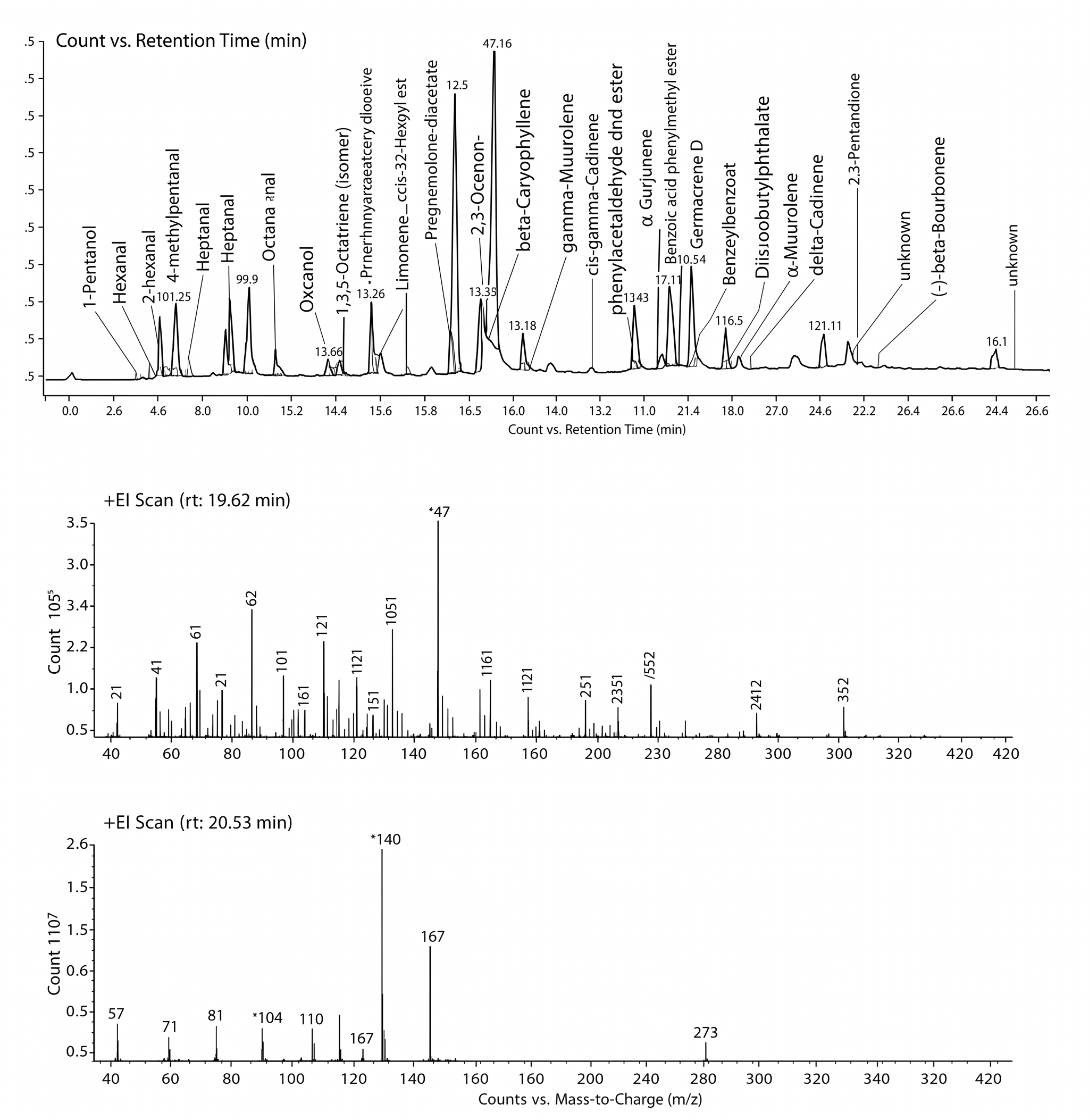
Gas chromatography–mass spectrometry (GC–MS) chromatogram of the ethanolic extract of Sargassum polycystum.
Major compounds identified by GC–MS in the ethanolic extract of
| Retention Time (min) | Compound Name | Chemical Class / Nature |
|---|---|---|
| 16.71 | Tetradecanoic acid (Myristic acid) | Saturated fatty acid |
| 17.66 | Hexadecanoic acid, methyl ester (Methyl palmitate) | Fatty acid methyl ester |
| 18.71 | Methyl stearate | Fatty acid methyl ester |
| 18.82 | Octadecanoic acid (Stearic acid) | Saturated fatty acid |
| 20.63 | Bis(2-ethylhexyl) phthalate | Ester compound |
HPLC Analysis
High-performance liquid chromatography (HPLC) analysis was conducted to characterize the principal photosynthetic pigments in the ethanolic extract of S. polycystum (Figure 2). The chromatogram displayed distinct, well-resolved peaks corresponding to chlorophylls and carotenoids (Table 3). Prominent peaks were identified as chlorophyll (RT = 1.45 min), pheophytin (RT = 1.63 min), xanthophyll (RT = 2.94 min), and fucoxanthin (RT = 3.53 min). The dominant peak at 2.94 min confirmed a high concentration of xanthophyll pigments in the algal matrix.
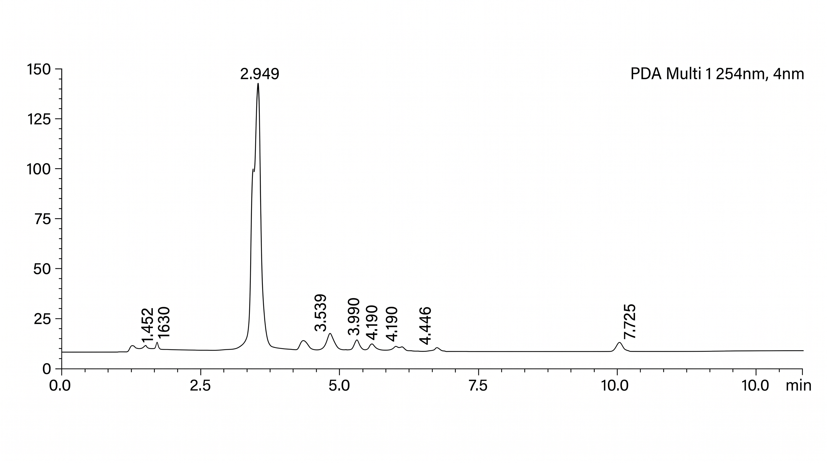
High-performance liquid chromatography (HPLC) chromatogram of the ethanolic extract of Sargassum polycystum.
Pigment composition identified by HPLC in the ethanolic extract of
| Retention Time (min) | Identified Compound |
|---|---|
| 1.45 | Chlorophyll |
| 1.63 | Pheophytin |
| 2.94 | Xanthophyll |
| 3.53 | Fucoxanthin |
Quantification of Total Phenolics and Flavonoids
Quantitative analysis revealed substantial amounts of polyphenolic compounds in the ethanolic extract of S. polycystum (Table 4). The total phenolic content was estimated at 2.13 ± 0.36 mg GAE g dw, while the total flavonoid content was 2.49 ± 0.42 mg RE g dw. The high recovery of these secondary metabolites highlights ethanol as an effective solvent for extracting polar antioxidant constituents from brown seaweeds.
Total phenolic and flavonoid contents of
| Parameter | Content (Mean ± SD, n = 3) |
|---|---|
| Total Phenolics | 2.13 ± 0.36 mg GAE g−1 dw |
| Total Flavonoids | 2.49 ± 0.42 mg RE g−1 dw |
Analysis of Pigment Composition
Thin-layer chromatography (TLC) of S. polycystum extracts prepared in solvents of varying polarity exhibited distinct pigment separation profiles (Table 5). The ethyl acetate extract yielded four distinct spots (Rf range 0.10–0.74), corresponding to neoxanthin and xanthophyll derivatives. Ethanol, acetone, and chloroform extracts showed individual bands corresponding to violaxanthin (Rf = 0.15), pheophytin (Rf = 0.80), and β-carotene (Rf = 0.84), respectively.
Thin-layer chromatography (TLC) separation of pigments in different solvent extracts of
| Solvent Extract | Number of Spots | Rf Value(s) | Tentatively Identified Pigment |
|---|---|---|---|
| Ethanol | 1 | 0.15 | Violaxanthin |
| Acetone | 1 | 0.80 | Pheophytin |
| Chloroform | 1 | 0.84 | β-Carotene |
| Ethyl acetate | 4 | 0.10–0.74 | Neoxanthin, Xanthophylls |
Spectrophotometric quantification confirmed substantial levels of photosynthetic pigments in both ethanolic and ethyl acetate extracts of S. polycystum (Table 6). The ethyl acetate extract exhibited higher total chlorophyll (7.89 ± 0.75 mg g), carotenoid (2.89 ± 0.55 mg g), and fucoxanthin (4.92 ± 0.68 mg g) concentrations compared to the ethanolic extract. Notably, fucoxanthin predominated over total carotenoids in both solvent extracts, underlining the pharmacological significance of this brown alga.
Spectrophotometric quantification of photosynthetic pigments in ethanol and ethyl acetate extracts of
| Solvent Extract | Chl | Chl | Total Chl (mg g−1) | Carotenoids (mg g−1) | Fucoxanthin (mg g−1) |
|---|---|---|---|---|---|
| Ethanol | 2.50 ± 0.39 | 5.16 ± 0.52 | 7.05 ± 0.62 | 1.86 ± 0.42 | 3.89 ± 0.64 |
| Ethyl acetate | 2.80 ± 0.46 | 5.78 ± 0.66 | 7.89 ± 0.75 | 2.89 ± 0.55 | 4.92 ± 0.68 |
Total Antioxidant Capacity
The total antioxidant capacity (TAC) of S. polycystum ethanolic extract increased in a concentration-dependent manner (Table 7). Absorbance values at 695 nm rose from 0.0040 ± 0.0008 at 100 µg/mL to 0.0200 ± 0.0012 at 500 µg/mL. Compared to the negative control (0.0020–0.0170), the extract exhibited consistently higher reducing capacity across all tested concentrations.
Total antioxidant capacity (TAC) of
| Concentration (µg/mL) | Control Absorbance (695 nm) | Ethanolic Extract Absorbance (695 nm) |
|---|---|---|
| 100 | 0.0020 ± 0.0005 | 0.0040 ± 0.0008 |
| 200 | 0.0060 ± 0.0007 | 0.0080 ± 0.0010 |
| 300 | 0.0080 ± 0.0010 | 0.0170 ± 0.0016 |
| 400 | 0.0120 ± 0.0010 | 0.0190 ± 0.0014 |
| 500 | 0.0170 ± 0.0011 | 0.0200 ± 0.0012 |
DPPH Radical Scavenging Activity
The ethanolic extract of S. polycystum demonstrated significant DPPH radical scavenging activity in a dose-dependent manner (Table 8). Radical inhibition increased from 14.12 ± 0.10% at 100 µg/mL to 83.57 ± 0.28% at 500 µg/mL. The calculated IC value for the ethanolic extract was 303.30 µg/mL, which was comparable to the standard ascorbic acid (IC = 289.54 µg/mL), demonstrating robust hydrogen-donating antioxidant efficiency.
DPPH radical scavenging potential of
| Concentration (µg/mL) | Ascorbic Acid (% Inhibition) | Ethanolic Extract (% Inhibition) |
|---|---|---|
| 100 | 23.25 ± 0.12 | 14.12 ± 0.10 |
| 200 | 41.36 ± 0.18 | 32.48 ± 0.15 |
| 300 | 58.72 ± 0.21 | 51.64 ± 0.19 |
| 400 | 74.85 ± 0.26 | 68.92 ± 0.23 |
| 500 | 88.41 ± 0.30 | 83.57 ± 0.28 |
| IC50 (µg/mL) | 289.54 | 303.30 |
ABTS Radical Scavenging Activity
Evaluation of ABTS radical cation scavenging confirmed the concentration-dependent antioxidant activity of the ethanolic extract (Table 9). Percentage inhibition rose from 18.64 ± 0.12% at 100 µg/mL to a maximum of 66.40 ± 0.27% at 500 µg/mL. The IC value of the extract was 347.60 µg/mL, compared with 237.29 µg/mL for standard ascorbic acid, reflecting moderate but substantial free-radical neutralization capacity.
ABTS radical scavenging potential of
| Concentration (µg/mL) | Ascorbic Acid (% Inhibition) | Ethanolic Extract (% Inhibition) |
|---|---|---|
| 100 | 34.82 ± 0.15 | 18.64 ± 0.12 |
| 200 | 52.47 ± 0.18 | 32.91 ± 0.16 |
| 300 | 68.75 ± 0.22 | 45.38 ± 0.19 |
| 400 | 81.96 ± 0.26 | 56.72 ± 0.23 |
| 500 | 92.51 ± 0.30 | 66.40 ± 0.27 |
| IC50 (µg/mL) | 237.29 | 347.60 |
HRBC Membrane Stabilization Anti-Inflammatory Assay
The HRBC membrane stabilization assay showed strong, concentration-dependent in vitro anti-inflammatory activity for the ethanolic extract of S. polycystum (Table 10). Membrane protection increased from 31.56 ± 0.15% at 100 µg/mL to 86.75 ± 0.27% at 500 µg/mL. At 500 µg/mL, the extract's protective efficacy surpassed that of the reference drug diclofenac sodium (85.43 ± 0.30%). The calculated IC value for the extract was 288.18 µg/mL (versus 251.72 µg/mL for diclofenac sodium), indicating effective erythrocyte membrane stabilization against hypotonic stress-induced hemolysis.
Human red blood cell (HRBC) membrane stabilization activity of
| Concentration (µg/mL) | Diclofenac Sodium (% Protection) | Ethanolic Extract (% Protection) |
|---|---|---|
| 100 | 38.24 ± 0.18 | 31.56 ± 0.15 |
| 200 | 55.68 ± 0.22 | 47.92 ± 0.19 |
| 300 | 69.84 ± 0.25 | 62.37 ± 0.21 |
| 400 | 80.26 ± 0.28 | 74.81 ± 0.24 |
| 500 | 85.43 ± 0.30 | 86.75 ± 0.27 |
| IC50 (µg/mL) | 251.72 | 288.18 |
In Vitro Antiproliferative Activity (MTT Assay)
The antiproliferative effect of S. polycystum ethanolic extract (SAR) on MCF-7 breast cancer cell viability was determined by the MTT assay. Exposure to SAR for 24 h induced a significant, dose-dependent decrease in MCF-7 cell survival (Figure 3). Cell viability decreased to 32.03% at 320 µg/mL, with an estimated IC value of 104.3 µg/mL. Microscopic examination revealed distinct morphological alterations in treated cells, including cell shrinkage, cytoplasmic condensation, membrane blebbing, and loss of cellular adherence, indicative of cytotoxic injury (Figure 4).
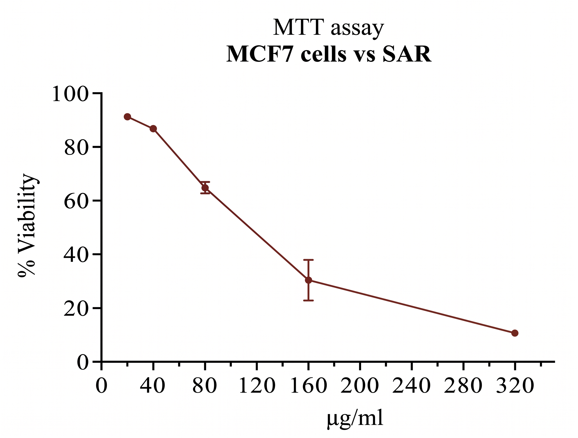
Effect of Sargassum polycystum ethanolic extract (SAR) on MCF-7 breast cancer cell viability measured by MTT assay after 24 h treatment.
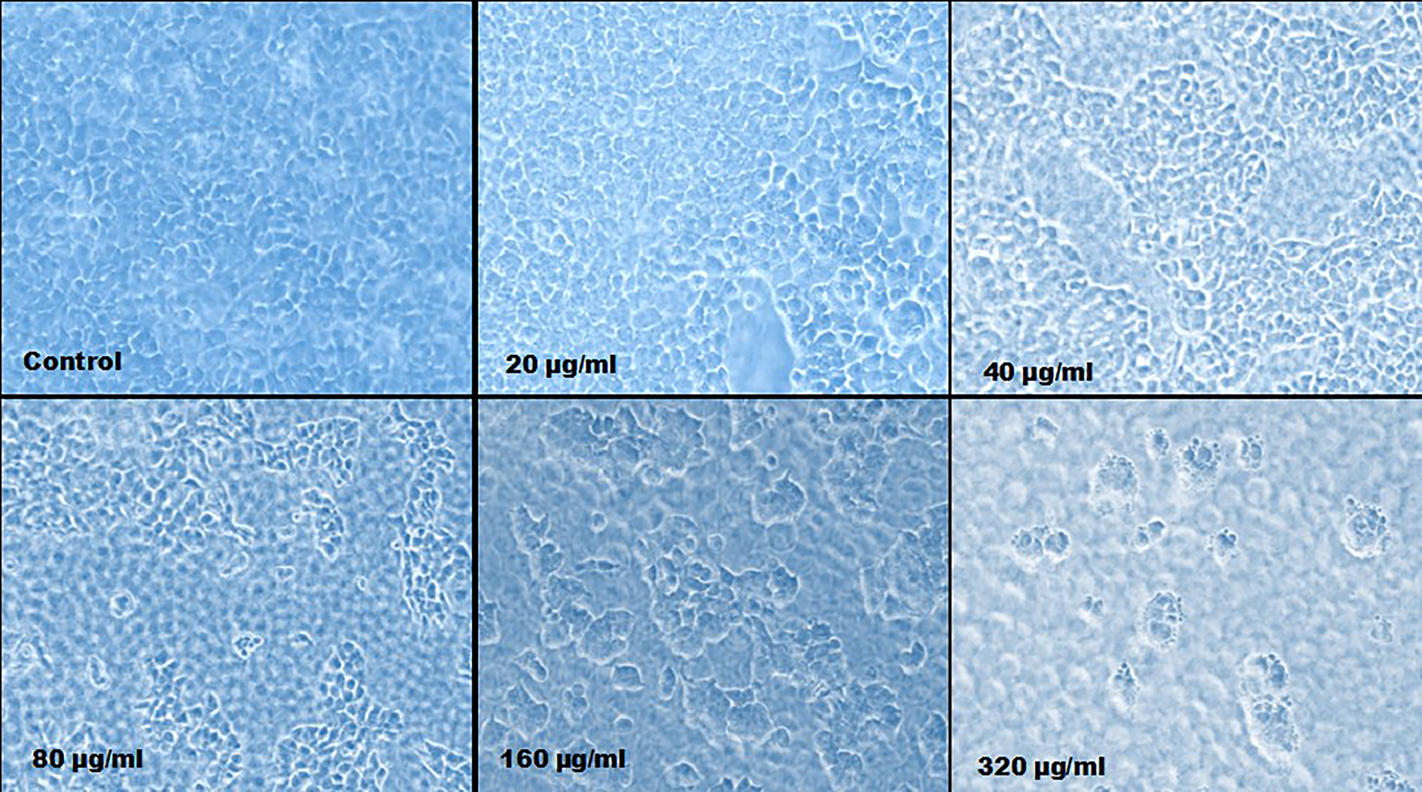
Morphological changes in MCF-7 human breast cancer cells observed under phase-contrast light microscopy following 24 h treatment with varying concentrations of Sargassum polycystum ethanolic extract.
DNA Fragmentation Analysis
Agarose gel electrophoresis of genomic DNA extracted from treated MCF-7 cells demonstrated a characteristic internucleosomal DNA ladder pattern (Figure 5). Untreated control cells (Lane 6) showed intact, high-molecular-weight genomic DNA without degradation. In contrast, cells treated with increasing concentrations of SAR (25, 50, and 100 µg/mL; Lanes 3–5) and Doxorubicin (positive control; Lane 2) exhibited dose-dependent DNA cleavage into oligonucleosomal fragments, confirming that SAR induces cell death via apoptosis rather than necrotic lysis.
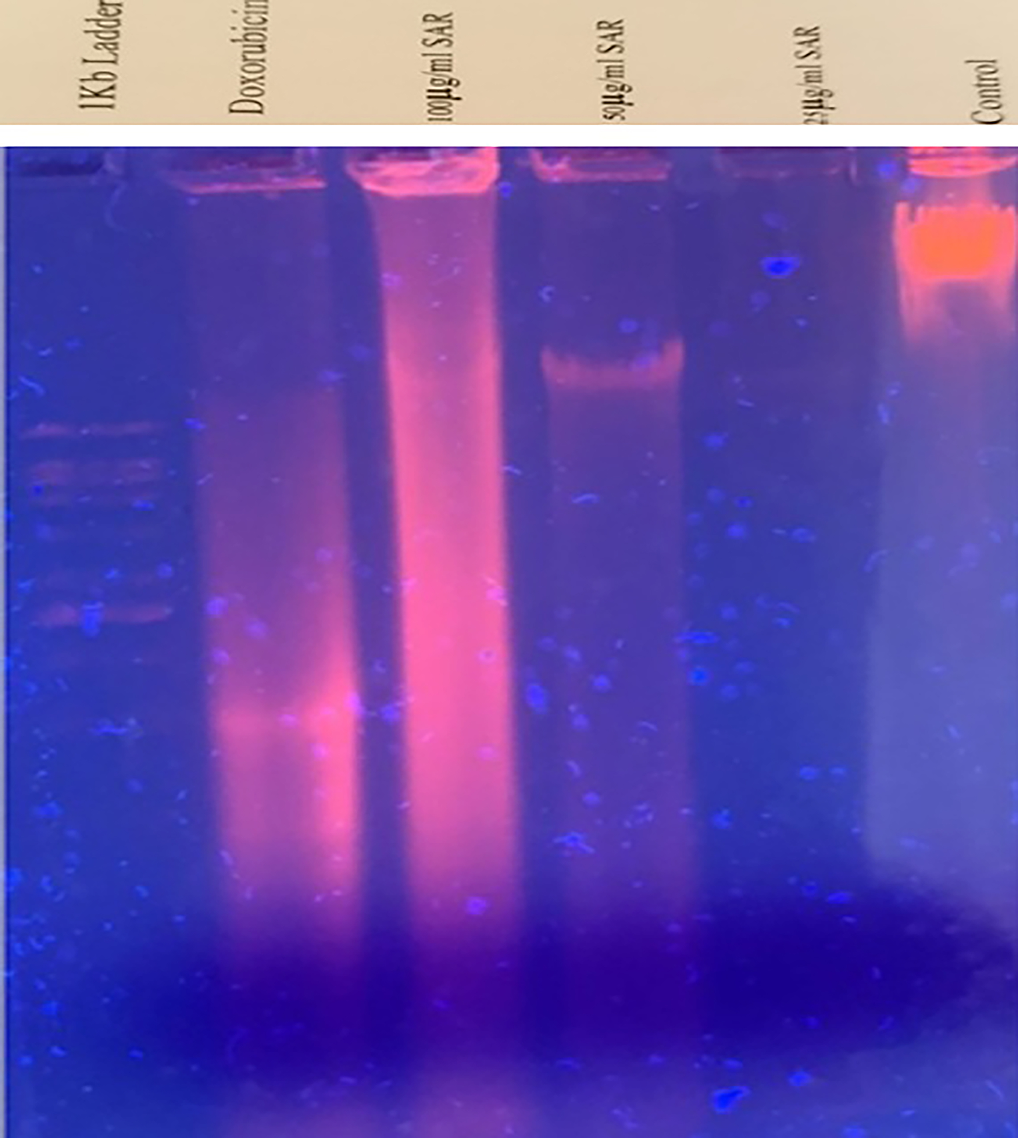
Agarose gel electrophoresis showing DNA fragmentation in MCF-7 cells after 24 h treatment. Lane 1: 1 kb DNA ladder; Lane 2: Doxorubicin (positive control); Lanes 3–5: MCF-7 treated with SAR at 25, 50, and 100 µg/mL, respectively; Lane 6: Untreated negative control.
Upregulation of p53 Gene Expression
To clarify the molecular signaling pathway of apoptosis, p53 mRNA expression in MCF-7 cells was quantified by RT-PCR (Figure 6). Treatment with 100 µg/mL SAR significantly upregulated p53 gene expression compared to untreated control cells, demonstrating a twofold increase in transcript level comparable to the positive control Doxorubicin. Specificity of PCR amplification was verified by a single sharp melting peak (Figure 7), indicating homogenous amplification. These findings confirm that S. polycystum triggers apoptosis in MCF-7 breast cancer cells via a p53-dependent pathway.
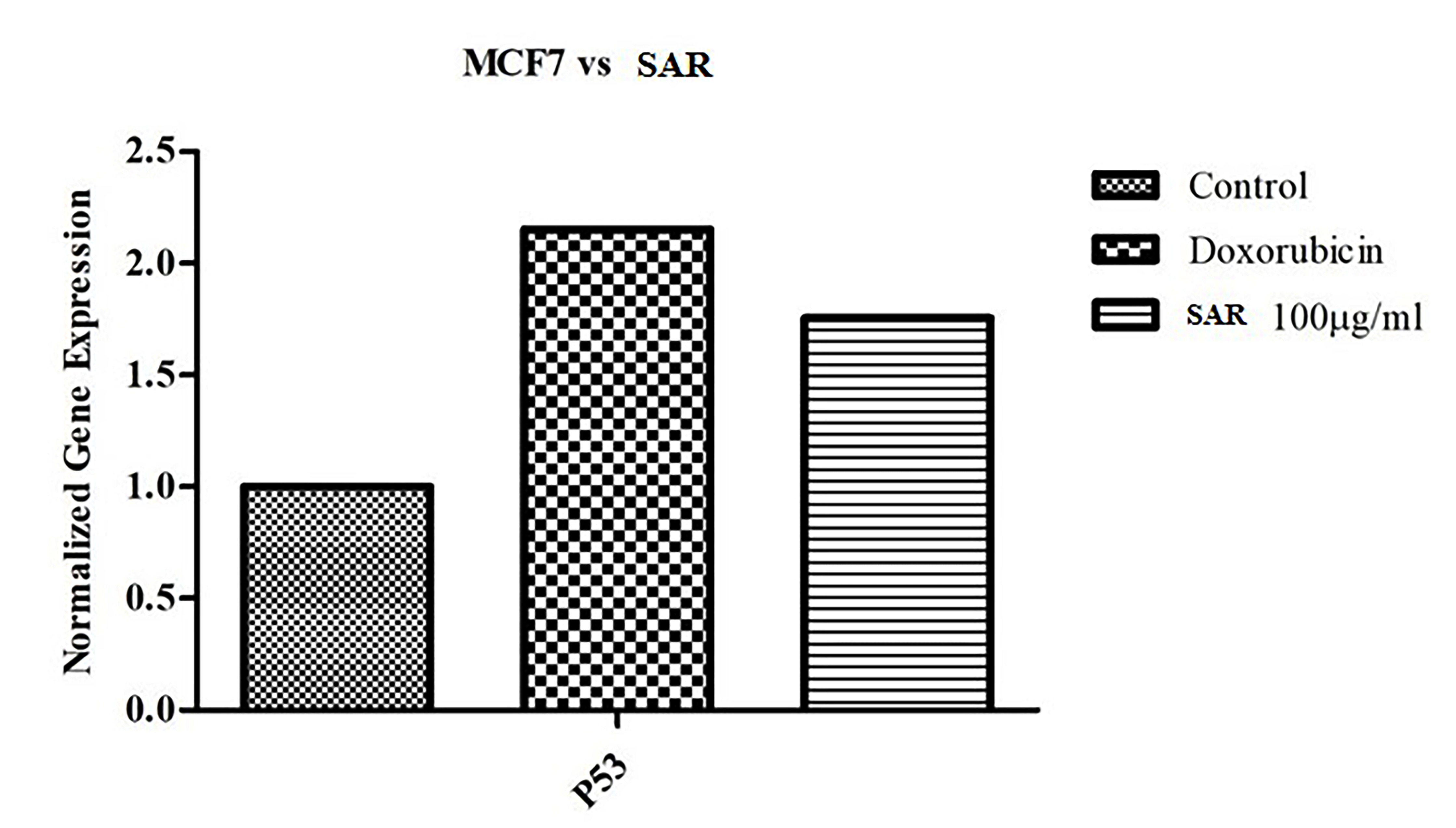
Effect of Sargassum polycystum ethanolic extract (SAR; 100 µg/mL) and Doxorubicin on
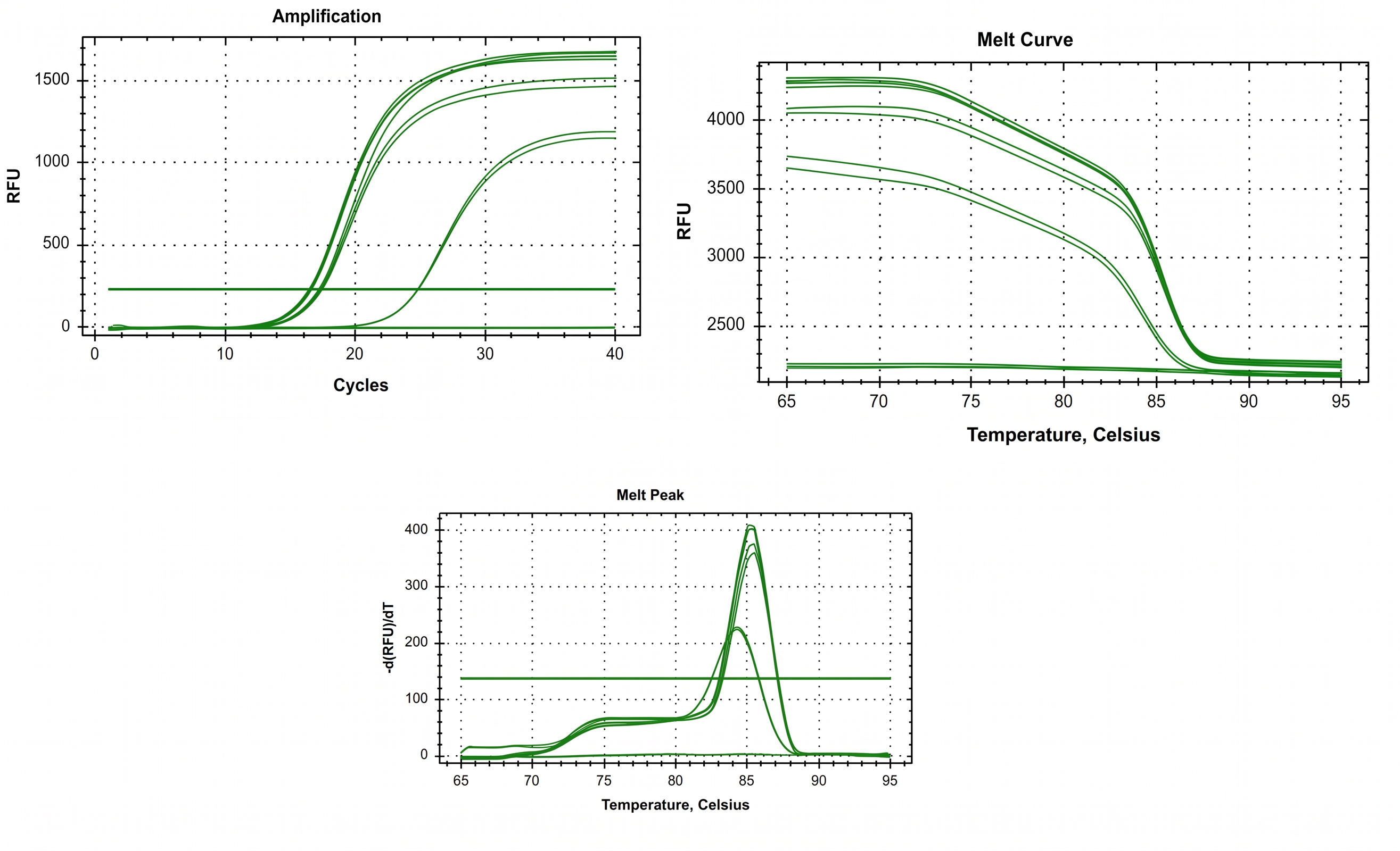
Amplification plot, melt curve, and melt peak analysis confirming specific, homogeneous RT-PCR amplification of the
DISCUSSION
Marine macroalgae represent rich, underutilized sources of secondary metabolites with high biomedical relevance. In the present investigation, ethanolic extract of Sargassum polycystum demonstrated significant antioxidant, anti-inflammatory, and antiproliferative activities in vitro, which are attributable to its rich chemical profile.
Qualitative phytochemical screening confirmed the presence of steroids, terpenoids, saponins, phenolics, flavonoids, tannins, and proteins, predominantly concentrated in the ethanolic extract. Phenolic compounds are established redox-active constituents capable of scavenging free radicals, chelating transition metal ions, and preventing lipid peroxidation30,13. The absence of alkaloids and anthraquinones suggests that the observed biological effects are primarily mediated by polyphenols, terpenoids, and photosynthetic pigments. HPLC and TLC profiling confirmed substantial concentrations of carotenoids, particularly fucoxanthin and xanthophylls, which are well-known for their strong antioxidant and cytoprotective capabilities31.
The antioxidant efficacy of S. polycystum was established across multiple complementary radical-scavenging assays. The total antioxidant capacity (TAC) assay demonstrated dose-dependent electron-donating reducing power24. Similarly, DPPH and ABTS assays confirmed potent free-radical neutralization, yielding IC values of 303.30 µg/mL and 347.60 µg/mL, respectively. These values align with previous studies on brown seaweeds, where moderate radical scavenging activities were reported due to complex crude mixtures of polyphenols, fucoxanthin, and sulfated polysaccharides32,33. Variations between our observed IC values and those reported in previous studies on S. polycystum by Arsianti et al. (298.32 µg/mL), Hidayati et al. (102.4 ± 0.056 µg/mL), and Gazali et al. (68.89 ± 5.36 µg/mL)34,35,36 can be attributed to environmental factors, geographical harvesting location, seasonal variations, and extraction methodologies.
Anti-inflammatory activity was corroborated using the HRBC membrane stabilization model. Lysosomal membrane stabilization is a crucial anti-inflammatory mechanism, as it prevents the extracellular release of hydrolytic enzymes and inflammatory mediators responsible for tissue injury28. Because the human erythrocyte membrane resembles the lysosomal membrane, the extract's ability to protect HRBCs against hypotonic lysis (86.75% protection at 500 µg/mL; IC = 288.18 µg/mL) strongly indicates membrane-stabilizing anti-inflammatory activity. This action may be linked to fatty acids (such as hexadecanoic and octadecanoic acids) and terpenoids detected by GC–MS, which inhibit inflammatory pathways and membrane disorganization.
Anticancer evaluation via the MTT assay demonstrated dose-dependent inhibition of MCF-7 breast cancer cell proliferation, yielding an IC value of 104.3 µg/mL. These results are consistent with previous investigations reporting cytotoxic effects of Sargassum species against carcinoma cell lines14,37, including S. cinctum extract against MCF-7 cells (IC = 134.50 µg/mL)38. Importantly, DNA fragmentation analysis revealed typical internucleosomal DNA laddering in agarose gel electrophoresis, confirming that SAR triggers regulated apoptotic cell death rather than acute necrotic lysis11.
Mechanistic exploration via RT-PCR established that SAR treatment induced a significant upregulation of the tumor suppressor gene p53 in MCF-7 cells. Tumor protein p53 regulates downstream effectors that control cell-cycle checkpoints, DNA repair, and pro-apoptotic gene transcription12. The observed upregulation of p53 correlates with reduced cell viability and DNA cleavage, confirming that S. polycystum extract promotes apoptosis through a p53-dependent signaling pathway.
Overall, these integrated findings highlight S. polycystum as a valuable marine resource of bioactive lead compounds. Because this study evaluated a crude ethanolic extract, the observed bioactivities likely result from additive or synergistic interactions among multiple secondary metabolites. Further research focusing on bioassay-guided fractionation, compound isolation, structural elucidation, and in vivo animal model validation is warranted to translate these findings into clinical applications.
CONCLUSION
The present study demonstrates that the ethanolic extract of Sargassum polycystum possesses potent antioxidant, anti-inflammatory, and in vitro antiproliferative activities against MCF-7 breast cancer cells. The extract inhibited cell viability, induced characteristic genomic DNA fragmentation, and activated p53-dependent apoptotic pathways. These findings provide compelling scientific support for the bioactivity of S. polycystum. Future investigations isolating active principles and elucidating in vivo therapeutic efficacy will be essential to advance its clinical development.
DECLARATIONS
Abbreviations
ABTS: 2,2'-azino-bis(3-ethylbenzothiazoline-6-sulfonic acid); ANOVA: Analysis of variance; ATCC: American Type Culture Collection; cDNA: Complementary deoxyribonucleic acid; Chl: Chlorophyll; DEPC: Diethyl pyrocarbonate; DMEM: Dulbecco's Modified Eagle Medium; DMSO: Dimethyl sulfoxide; DNA: Deoxyribonucleic acid; DPPH: 2,2-diphenyl-1-picrylhydrazyl; dw: Dry weight; EDTA: Ethylenediaminetetraacetic acid; FBS: Fetal bovine serum; GAE: Gallic acid equivalents; GC–MS: Gas chromatography–mass spectrometry; HPLC: High-performance liquid chromatography; HRBC: Human red blood cell; IC: Half-maximal inhibitory concentration; MCF-7: Michigan Cancer Foundation-7; MTT: 3-(4,5-dimethylthiazol-2-yl)-2,5-diphenyltetrazolium bromide; NSAID: Non-steroidal anti-inflammatory drug; OD: Optical density; ORCID: Open Researcher and Contributor ID; p53: Tumor protein p53; PBS: Phosphate-buffered saline; PCR: Polymerase chain reaction; RE: Rutin equivalents; Rf: Retention factor; RNA: Ribonucleic acid; ROS: Reactive oxygen species; RT-PCR: Reverse transcription-polymerase chain reaction; SAR: Sargassum polycystum ethanolic extract; SD: Standard deviation; TAC: Total antioxidant capacity; TFC: Total flavonoid content; TLC: Thin-layer chromatography; TPC: Total phenolic content; UV: Ultraviolet.
Acknowledgments
The authors express their sincere gratitude to the management of Mohamed Sathak College of Arts and Science, Greater Chennai, Tamil Nadu, India, for providing the necessary research facilities and laboratory infrastructure to carry out this work.
Author’s contributions
Conceptualization and study design: SAMY and AV; Experimental execution and investigation: VK; Data analysis and software/data curation: JSP and YN; Writing – original draft preparation, review, and editing: All authors. All authors read and approved the final manuscript.
Funding
This research received no specific grant or financial support from any funding agency in the public, commercial, or non-profit sectors.
Availability of data and materials
All data generated or analyzed during this study are included in this published article, and further details or raw materials are available from the corresponding author upon reasonable request.
Ethics approval and consent to participate
Human red blood cells were collected from a healthy human volunteer solely for the in vitro HRBC membrane stabilization assay in accordance with institutional guidelines and following informed consent. For the in vitro cell line studies, ethical approval was not required.
Consent for publication
Not applicable.
Declaration of generative AI and AI-assisted technologies in the writing process
The authors declare that no generative AI or AI-assisted technologies were used in the writing process or preparation of this manuscript.
Competing interests
The authors declare that they have no competing interests.