


Survival Profiles and Predictive Factors of Acral Melanoma in Viet Nam: A 10-Year Single-Center Retrospective Cohort Study
- Department of Dermatology, Hanoi Medical University, Hanoi, Vietnam
- Department of Plastic, Aesthetic Surgery and Rehabilitation, National Hospital of Dermatology and Venereology, Hanoi, Vietnam
- Board of Directors, National Hospital of Dermatology and Venereology, Hanoi, Vietnam
- Department of Pathology, National Hospital of Dermatology and Venereology, Hanoi, Vietnam
Abstract
Introduction: Acral melanoma (AM) is the most prevalent melanoma subtype in Asian populations; however, long-term survival data from Southeast Asia, particularly Vietnam, remain scarce. This study aimed to investigate the clinicopathological characteristics and prognostic factors influencing overall survival (OS) in a Vietnamese cohort of patients with AM.
Methods: A retrospective cohort study was conducted on 84 adult patients who underwent surgical resection for primary AM at a single tertiary referral center between January 2016 and January 2026. Clinicopathological correlations were evaluated, and OS was analyzed using the Kaplan-Meier method and multivariable Cox proportional hazards regression modeling.
Results: The mean patient age was 59.49 ± 13.79 years, with a slight female predominance (54.8%). Most patients presented at advanced clinical stages (65.5% Stage II/III), with 60.7% exhibiting macroscopic ulceration. The heel pad was the most common primary site (35.7%). Regional lymph node metastasis was present in 16.7% of cases at diagnosis. Over a mean follow-up of 40.17 months, the all-cause mortality rate was 22.6%. Univariate log-rank analysis identified AJCC clinical stage (p = 0.041), regional lymph node status (p = 0.029), and macroscopic ulceration (p = 0.027) as significant prognostic factors for OS. In the multivariable Cox model, both regional lymph node metastasis (Hazard Ratio [HR] = 4.735; 95% CI: 1.666–13.458; p = 0.004) and Breslow thickness ≥ 2.0 mm (HR = 3.155; 95% CI: 1.007–9.885; p = 0.049) emerged as independent predictors of decreased overall survival.
Conclusions: Patients with AM in Vietnam frequently present with advanced, thick, and ulcerated primary tumors. Regional lymph node metastasis and Breslow thickness are the dominant independent determinants of survival, highlighting the critical need for public health initiatives focused on early detection and standardized nodal evaluation.
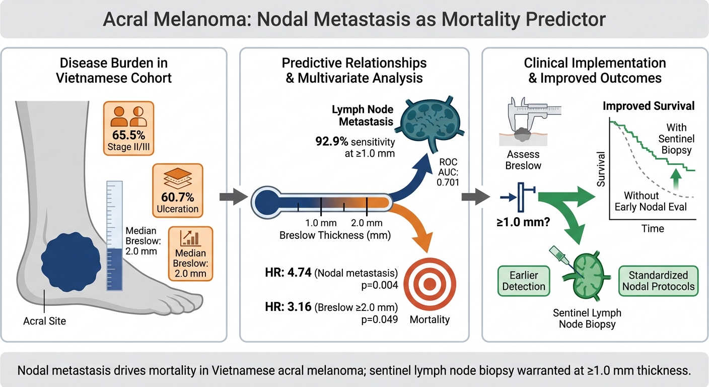
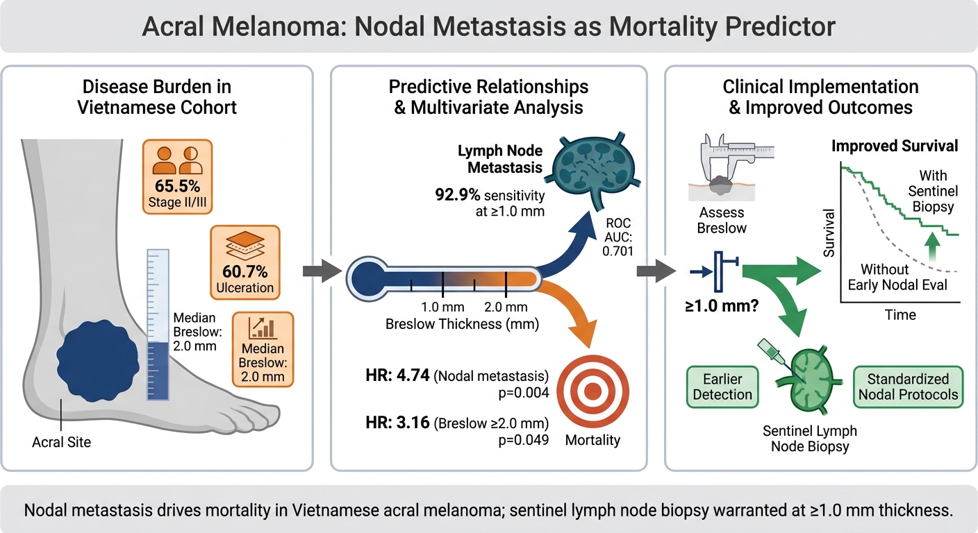
Graphical abstract
INTRODUCTION
The global incidence of cutaneous melanoma is steadily rising, a trend that encompasses acral melanoma (AM)—a distinct clinical and biological entity arising on the non-hair-bearing, glabrous skin of the palms, soles, and nail apparatus1,2. Although AM accounts for only 2% to 3% of all cutaneous melanomas in Western populations, it constitutes the most predominant subtype among non-Caucasian individuals. Strikingly, across several East and Southeast Asian populations—including Taiwan, China, Japan, Korea, Hong Kong, and Singapore—AM represents up to 58% of all diagnosed melanoma cases3,4. Beyond its unique epidemiological profile, AM possesses a distinct clinical, genomic, and microenvironmental landscape compared with melanomas originating from non-glabrous cutaneous sites2. While non-acral cutaneous melanomas are predominantly driven by ultraviolet (UV)-induced DNA damage and BRAF mutations, AM exhibits a lower mutational burden characterized by complex structural variants, focal gene amplifications, and recurrent alterations within the KIT, NRAS, and MAP-kinase signaling pathways5.
Clinically, AM is notoriously associated with delayed diagnosis and an aggressive disease trajectory. European and American registry data reflect this disparity, demonstrating that 62% of AM cases are diagnosed at Stage II or higher, compared with only 32% of non-acral cutaneous melanomas2. Consequently, the prognostic outlook for AM is substantially worse, as evidenced by a significantly lower 5-year melanoma-specific survival (MSS) rate relative to non-acral cutaneous melanoma (80.6% vs. 93.0%)6. Despite the disproportionately high burden of AM in Asian populations, current evidence regarding its prognostic determinants is largely derived from cohorts in East Asia or Western countries. Real-world data from Southeast Asia, particularly Vietnam, remain exceptionally limited. In these resource-constrained settings, patients with AM frequently present at advanced stages with deep vertical tumor invasion and a high prevalence of macroscopic ulceration. This delayed presentation is largely attributed to the concealed anatomical locations of primary lesions, atypical initial clinical presentations, and limited public awareness. Thus, clarifying the independent prognostic impacts of primary tumor characteristics—such as Breslow thickness and ulceration—versus regional lymph node involvement within this specific demographic context is clinically imperative.
To address this crucial geographic and literature gap, the present study investigated the clinicopathological characteristics and long-term overall survival (OS) outcomes of an adult AM cohort treated surgically over a 10-year period at a national tertiary referral center in Vietnam. Furthermore, using multivariable Cox regression analysis to rigorously adjust for clinical confounders such as patient age and underlying comorbidities, we aimed to identify independent predictors of OS. These real-world data from Vietnam provide valuable insights into the global epidemiological spectrum of AM and offer evidence to guide clinical risk stratification and management strategies.
MATERIALS AND METHODS
Study Design and Ethical Approval
This single-center, retrospective cohort study was conducted at the National Hospital of Dermatology and Venereology (Hanoi, Vietnam). The study protocol was reviewed and formally approved by the Institutional Review Board (IRB) and Ethics Committee of the National Hospital of Dermatology and Venereology (Approval Decision No. 2869/QĐ-BVDLTW, dated October 23, 2025). Under this IRB decision, the requirement for individual written informed consent was waived due to the retrospective study design and the use of de-identified clinical data. All study procedures were performed in strict compliance with the ethical principles of the Declaration of Helsinki (1964) and its later amendments. Reporting adheres to the Strengthening the Reporting of Observational Studies in Epidemiology (STROBE) statement guidelines.
Materials, Study Population, and Data Sources
Electronic medical records, surgical logs, and archived formalin-fixed paraffin-embedded (FFPE) histopathological specimens were systematically screened for adult patients (aged ≥ 18 years) who underwent primary surgical treatment for histologically confirmed AM between January 2016 and January 2026. A total of 118 patient records with a primary diagnosis of melanoma were initially identified. Thirty-four patients were excluded based on predefined criteria: 15 patients with non-acral cutaneous melanoma subtypes; 11 patients lost to follow-up before reaching the required evaluation timeframe; and 8 patients with missing key clinical or histopathological data. Ultimately, a final cohort of 84 patients met all inclusion criteria and was included in the definitive survival analysis.
Clinical and Histopathological Data Extraction
Demographic and clinical data were collected using a standardized case report form, including age at diagnosis, gender, presence of underlying medical comorbidities, anatomical subsite of the primary lesion (categorized as plantar, palmar, or subungual/nail unit), duration of symptoms prior to surgical intervention, and macroscopic ulceration status. Histopathological slides were independently evaluated by experienced dermatopathologists. Because the 10-year study timeframe (2016–2026) spanned the transition between the 7th and 8th editions of the American Joint Committee on Cancer (AJCC) staging systems, all archived FFPE tissue blocks were retrieved, re-sectioned, and re-stained with hematoxylin and eosin (H&E). Primary tumor characteristics, specifically Breslow thickness and microscopic ulceration, were re-evaluated, and all cases were uniformly re-staged according to the AJCC 8th Edition guidelines to ensure staging consistency across the cohort. The primary study endpoint was overall survival (OS), calculated from the date of primary surgical resection to the date of death from any cause or the date of last clinical follow-up.
Statistical Analysis
Statistical analyses were performed using IBM SPSS Statistics for Windows, Version 27.0 (IBM Corp., Armonk, NY, USA). Continuous variables are expressed as mean ± standard deviation (SD) or median (range/interquartile range [IQR]), depending on normality of distribution. Categorical variables are presented as absolute counts (n) and percentages (%). Associations between clinicopathological parameters and regional lymph node metastasis were evaluated using Pearson's Chi-square test or Fisher's exact test, as appropriate. Receiver operating characteristic (ROC) curve analysis was performed to determine the predictive capacity of Breslow thickness for regional lymph node metastasis and to identify the optimal clinical diagnostic threshold. OS probabilities were generated using the Kaplan-Meier method, and differences between survival curves were assessed using the log-rank test. To identify independent prognostic factors for OS while adjusting for potential clinical confounders, a multivariable Cox proportional hazards regression model was constructed. The proportional hazards assumption was assessed and validated for all model covariates using Schoenfeld residual diagnostics. To prevent multicollinearity, the composite AJCC clinical stage was omitted from the multivariable model, as it is directly derived from Breslow thickness and nodal status. Multicollinearity among remaining predictors was assessed using the Variance Inflation Factor (VIF), with all VIF values remaining well below the threshold of 5.0. Post-hoc power analysis confirmed that the final sample size (n = 84) provided adequate statistical power (> 80%) for the primary multivariable Cox regression hazard ratios. All statistical tests were two-tailed, and a p-value < 0.05 was considered statistically significant.
RESULTS
Baseline Demographics and Clinicopathological Characteristics
A total of 84 patients with histologically confirmed acral melanoma (AM) were analyzed. Baseline clinicopathological characteristics are summarized in Table 1. The mean patient age was 59.49 ± 13.79 years (range: 22–89 years), with a slight female predominance (54.8%, n = 46). Underlying medical comorbidities were documented in 40.5% (n = 34) of the cohort. The plantar surface was the most frequent anatomical subsite, primarily affecting the heel pad (35.7%, n = 30), followed by the base of the 5th metatarsal (14.3%, n = 12) and the medial longitudinal arch (14.3%, n = 12). Palmar and subungual lesions together accounted for 19.0% (n = 16) of cases. Marked diagnostic delay was evident, with 79.7% (n = 67) of patients reporting a disease duration exceeding 1 year prior to definitive surgery (median duration: 24 months). Consequently, primary tumors presented with advanced local features: macroscopic ulceration was present in 60.7% (n = 51) of cases, and 42.9% (n = 36) had a Breslow thickness ≥ 2.0 mm. The median Breslow thickness was 2.0 mm (mean: 2.66 ± 2.41 mm; range: 0.0–12.0 mm). At initial presentation, 16.7% (n = 14) of patients had established regional lymph node metastasis, and 65.5% (n = 55) presented at advanced clinical stages (Stage II or III).
Baseline Demographics and Clinicopathological Characteristics of Patients with Acral Melanoma (n = 84). Demographic, clinical, and histopathological features of 84 adult patients with acral melanoma (AM) who underwent primary surgical resection at the National Hospital of Dermatology and Venereology (Hanoi, Vietnam) between January 2016 and January 2026. Continuous variables are expressed as mean ± standard deviation (SD) or median (range), while categorical variables are reported as frequencies (n) and percentages (%). Tumor staging was evaluated according to the American Joint Committee on Cancer (AJCC) 8th Edition criteria. Abbreviations: AJCC, American Joint Committee on Cancer; SD, standard deviation.
| Characteristic | Category / Parameter | Overall Cohort (n = 84) |
|---|---|---|
| Age (years) | Mean ± SD | 59.49 ± 13.79 |
| Age (years) | Median (Range) | 60.5 (22–89) |
| Gender, n (%) | Female | 46 (54.8%) |
| Male | 38 (45.2%) | |
| Underlying Comorbidities, n (%) | Present | 34 (40.5%) |
| Absent | 50 (59.5%) | |
| Primary Tumor Subsite, n (%) | Heel pad | 30 (35.7%) |
| Base of 5th metatarsal | 12 (14.3%) | |
| Medial longitudinal arch | 12 (14.3%) | |
| Other plantar sites | 14 (16.7%) | |
| Palmar / Subungual | 16 (19.0%) | |
| Disease Duration Prior to Surgery, n (%) | > 1 year | 67 (79.7%) |
| ≤ 1 year | 17 (20.3%) | |
| Macroscopic Ulceration, n (%) | Present | 51 (60.7%) |
| Absent | 33 (39.3%) | |
| Breslow Thickness (mm) | Median (Range) | 2.0 (0.0–12.0) |
| Mean ± SD | 2.66 ± 2.41 | |
| < 2.0 mm | 48 (57.1%) | |
| ≥ 2.0 mm | 36 (42.9%) | |
| Regional Lymph Node Status, n (%) | Metastasis present | 14 (16.7%) |
| Metastasis absent | 70 (83.3%) | |
| AJCC Clinical Stage (8th Ed.), n (%) | Stage I–II (Early) | 29 (34.5%) |
| Stage III–IV (Advanced) | 55 (65.5%) |
Predictors of Regional Lymph Node Metastasis
Univariate cross-tabulation analysis (Table 2) demonstrated a statistically significant association between primary tumor Breslow thickness and regional lymph node metastasis (p = 0.023). In contrast, disease duration (p = 0.849), macroscopic ulceration (p = 0.369), age group (p = 0.400), gender (p = 0.170), and primary anatomical site (p = 1.000) showed no statistically significant association with nodal status. ROC curve analysis further confirmed that Breslow thickness significantly discriminated between patients with and without regional lymph node metastasis, yielding an area under the curve (AUC) of 0.701 (95% CI: 0.542–0.859; p = 0.018) (Figure 1). Coordinate analysis established that a Breslow thickness threshold of ≥ 1.0 mm optimized diagnostic sensitivity at 92.9% for identifying nodal involvement in this cohort.
Associations Between Clinicopathological Variables and Regional Lymph Node Metastasis in Acral Melanoma (n = 84). Univariate cross-tabulation analysis of clinicopathological characteristics associated with regional lymph node metastasis in adult patients with acral melanoma (n = 84). Statistical comparisons between categorical variables were performed using Pearson's Chi-square test or Fisher's exact test, as appropriate. Two-tailed p-values < 0.05 indicate statistical significance (*). Breslow thickness demonstrated a significant association with nodal invasion (p = 0.023). Abbreviations: AJCC, American Joint Committee on Cancer; n, number of cases.
| Variable | Category | Lymph Node Negative (n = 70) | Lymph Node Positive (n = 14) | p-value |
|---|---|---|---|---|
| Breslow Thickness | < 1.0 mm | 17 (94.4%) | 1 (5.6%) | 0.023* |
| 1.0 - < 2.0 mm | 26 (86.7%) | 4 (13.3%) | 0.023* | |
| ≥ 2.0 mm | 27 (75.0%) | 9 (25.0%) | 0.023* | |
| Macroscopic Ulceration | Absent | 29 (87.9%) | 4 (12.1%) | 0.369 |
| Present | 41 (80.4%) | 10 (19.6%) | 0.369 | |
| Disease Duration | ≤ 1 year | 14 (82.4%) | 3 (17.6%) | 0.849 |
| > 1 year | 56 (83.6%) | 11 (16.4%) | 0.849 | |
| Age Group | < 60 years | 31 (86.1%) | 5 (13.9%) | 0.400 |
| ≥ 60 years | 39 (81.25%) | 9 (18.75%) | 0.400 | |
| Gender | Female | 41 (89.1%) | 5 (10.9%) | 0.170 |
| Male | 29 (76.3%) | 9 (23.7%) | 0.170 | |
| Anatomical Subsite | Plantar | 45 (83.3%) | 9 (16.7%) | 1.000 |
| Non-plantar (Palmar/Subungual) | 25 (83.3%) | 5 (16.7%) | 1.000 |
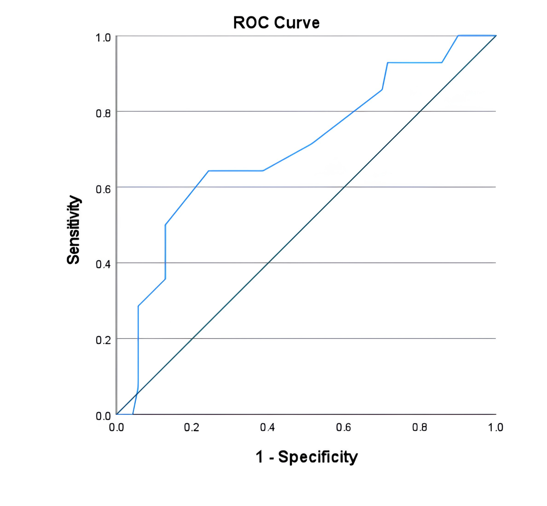
Predictive capacity of primary tumor Breslow thickness for regional lymph node metastasis in acral melanoma. Receiver operating characteristic (ROC) curve demonstrating that primary tumor Breslow thickness significantly predicts regional lymph node metastasis in acral melanoma (n = 84). ROC analysis was performed to quantify the discriminative ability of tumor depth for occult nodal involvement. The area under the ROC curve (AUC) is 0.701 (95% CI: 0.542–0.859; p = 0.018). Coordinate analysis established an optimal clinical decision cutoff at Breslow thickness ≥ 1.0 mm, yielding a sensitivity of 92.9% and a negative predictive value of 94.4% for ruling out regional lymph node metastasis. The diagonal reference line represents chance performance (AUC = 0.50).
Univariate Survival Analysis
Over a mean follow-up period of 40.17 ± 33.53 months (range: 1–114 months), 19 all-cause deaths (22.6%) were documented. Unadjusted Kaplan-Meier survival analysis (Figure 2) revealed several significant baseline predictors of decreased overall survival. Patients presenting with advanced AJCC clinical stage (Stage III–IV) had significantly worse OS compared with those presenting with early-stage disease (Stage I–II; log-rank p = 0.041; Figure 2D). Similarly, the presence of regional lymph node metastasis (log-rank p = 0.029; Figure 2G) and macroscopic tumor ulceration (log-rank p = 0.027; Figure 2C) significantly reduced OS. Conversely, no statistically significant survival differences were observed based on gender (p = 0.789; Figure 2A), primary anatomical subsite (p = 1.000; Figure 2B), age group (p = 0.083), or unadjusted Breslow thickness categorizations (4-tier log-rank p = 0.757, Figure 2E; 2-tier log-rank p = 0.852, Figure 2F).
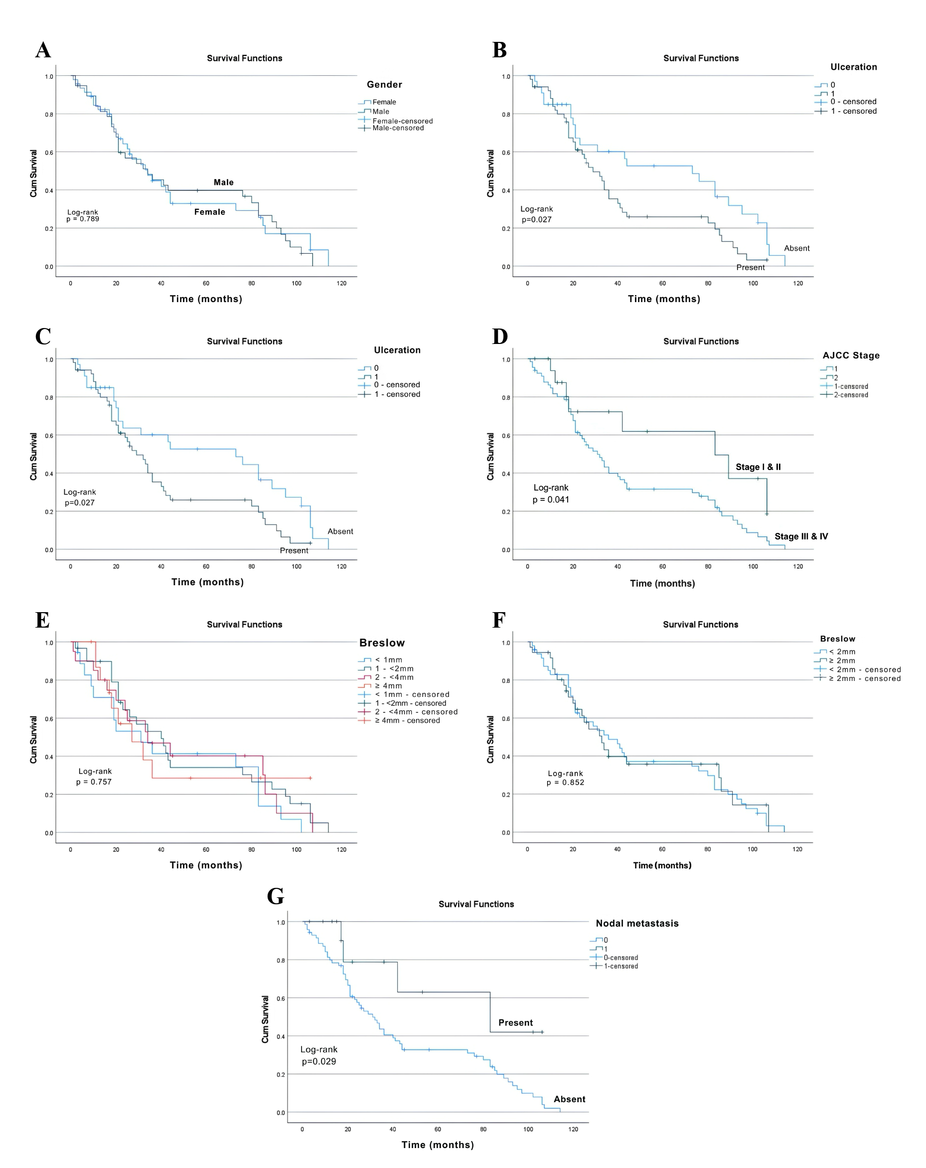
Kaplan-Meier overall survival estimates for patients with acral melanoma stratified by clinical and histopathological parameters (n = 84). Unadjusted Kaplan-Meier survival curves illustrating overall survival (OS) probabilities in acral melanoma according to key baseline parameters over a mean follow-up period of 40.17 months. (A) OS stratified by Gender: Comparison between male (n = 38) and female (n = 46) patients shows no statistically significant survival difference (log-rank p = 0.789). (B) OS stratified by Anatomical Subsite: OS curves comparing plantar (n = 54), palmar (n = 10), and subungual (n = 20) primary tumor sites demonstrate comparable survival outcomes (log-rank p = 1.000). (C) OS stratified by Macroscopic Tumor Ulceration: Patients presenting with ulcerated tumors (n = 51) exhibit significantly lower OS compared to those without ulceration (n = 33; log-rank p = 0.027). (D) OS stratified by AJCC Clinical Stage: Advanced clinical stage (Stage III–IV, n = 55) is associated with significantly reduced OS compared to early-stage disease (Stage I–II, n = 29; log-rank p = 0.041). (E) OS stratified by 4-Tier Breslow Thickness: Categorization into four depth tiers (< 1.0 mm, 1.0 - < 2.0 mm, 2.0 - 4.0 mm, and > 4.0 mm) shows no significant unadjusted survival distinction (log-rank p = 0.757). (F) OS stratified by 2-Tier Breslow Thickness: Dichotomization at 2.0 mm (< 2.0 mm vs. ≥ 2.0 mm) similarly shows no unadjusted survival divergence (log-rank p = 0.852). (G) OS stratified by Regional Lymph Node Status: The presence of regional lymph node metastasis (n = 14) significantly impairs OS compared to nodal-negative disease (n = 70; log-rank p = 0.029). Cumulative survival probabilities were estimated using the Kaplan-Meier method, and differences between survival curves were evaluated using the two-tailed log-rank test (p < 0.05 considered statistically significant).
Independent Prognostic Factors for Overall Survival
To evaluate the independent prognostic impact of primary tumor parameters while controlling for potential clinical confounders, a multivariable Cox proportional hazards regression model was performed (Figure 3). After adjusting for patient age, macroscopic tumor ulceration, medical comorbidities, and Breslow thickness, regional lymph node metastasis remained the strongest independent predictor of all-cause mortality, conferring a nearly five-fold increase in death hazard (Hazard Ratio [HR] = 4.735; 95% CI: 1.666–13.458; p = 0.004; Figure 3A). Furthermore, multivariable adjustment unmasked primary tumor invasion depth as an independent prognostic factor: patients with a Breslow thickness ≥ 2.0 mm demonstrated a significantly higher mortality hazard compared with those with thinner lesions (< 2.0 mm) (HR = 3.155; 95% CI: 1.007–9.885; p = 0.049; Figure 3B).
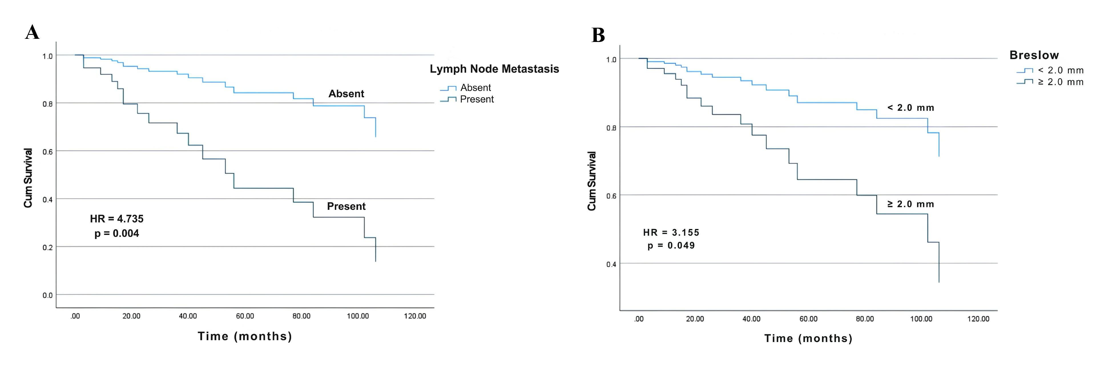
Covariate-adjusted overall survival curves derived from multivariate Cox proportional hazards regression modelling (n = 84). Multivariate Cox proportional hazards survival curves revealing regional lymph node metastasis and Breslow thickness as independent predictors of all-cause mortality in acral melanoma, after adjusting for clinical confounders. Survival curves reflect multivariate model predictions after controlling for age group, macroscopic tumor ulceration, and underlying comorbidities. (A) Adjusted OS by Regional Lymph Node Metastasis Status: Nodal involvement is the strongest independent predictor of poor overall survival, conferring a nearly five-fold increase in death hazard (Hazard Ratio [HR] = 4.735; 95% CI: 1.666–13.458; p = 0.004). (B) Adjusted OS by Primary Tumor Breslow Thickness: Accounting for clinical confounders unmasks primary tumor depth (≥ 2.0 mm vs. < 2.0 mm) as a potent independent prognostic factor, associated with a more than three-fold elevated risk of death (HR = 3.155; 95% CI: 1.007–9.885; p = 0.049). Multivariable modeling was conducted using Cox proportional hazards regression. Proportional hazards assumptions were confirmed via Schoenfeld residual testing.
DISCUSSION
This 10-year retrospective cohort study elucidates the clinical presentation, prognostic determinants, and survival outcomes of acral melanoma (AM) in a Vietnamese population. Our findings highlight a profound pattern of diagnostic delay, characterized by thick primary tumors, high rates of macroscopic ulceration, and frequent presentation at advanced clinical stages. Crucially, multivariable Cox regression analysis established regional lymph node metastasis and Breslow thickness as independent predictors of overall survival in this Southeast Asian cohort.
Unlike non-acral cutaneous melanoma, which is primarily initiated by UV radiation-induced mutagenesis, AM develops along a distinct oncogenic trajectory. Pathogenetically, AM tumorigenesis involves early genomic instability—often driven by telomere crisis or mechanical stress—leading to structural chromosomal variations and gene copy number alterations ("hailstorm" patterns), followed by TERT promoter activation and late-stage MAP-kinase signaling pathway mutations5. Clinically, this aggressive biology is exacerbated by substantial diagnostic delays. In our cohort, nearly 80% of patients experienced symptoms for over 1 year prior to surgical resection (median duration: 24 months), culminating in 65.5% presenting at advanced stages (Stage II/III) and 42.9% exhibiting a Breslow thickness ≥ 2.0 mm. Deep vertical invasion in AM has been shown to inversely correlate with protective immune cell infiltration (such as tumor-infiltrating lymphocytes and CD3+/CD4+ T cells) while promoting an immunosuppressive tumor microenvironment enriched with CD68+ tumor-associated macrophages7. Furthermore, thick AM lesions frequently harbor higher rates of copy number variations (CNVs) and structural variants in NRAS or "Triple Wild-Type" genomic backgrounds, whereas BRAF mutations are less frequent in deeper lesions (p = 0.02)8. These molecular features explain the aggressive disease course observed in thick AM primary tumors.
The predominant anatomical distribution observed in our cohort—specifically weight-bearing plantar regions, such as the heel pad (35.7%)—further highlights the role of mechanical stress in disease progression. Aside from concealing early lesions from self-examination, weight-bearing surfaces are subjected to continuous mechanical strain during ambulation. Mechanobiological studies suggest that cyclic mechanical forces activate YAP transcription factor signaling at the invasive tumor front, causing nuclear envelope disruption, genomic instability, and activation of cGAS-STING inflammatory signaling pathways that promote local invasion and metastatic dissemination9.
The high prevalence of macroscopic ulceration in our cohort (60.7%) aligns with reports from other developing regions and reflects both delayed clinical presentation and intrinsic tumor aggressiveness10,11. Primary tumor depth and ulceration are tightly associated with genomic instability, total CNV burden, and localized immunosuppression8. These mechanisms consistent with our univariate findings, wherein advanced AJCC stage, tumor ulceration, and regional nodal metastasis were significantly associated with reduced overall survival. Interestingly, although subungual melanomas display distinct molecular profiles compared with volar lesions (e.g., lower rates of BRAF mutations [0% vs. 13%] and lower objective response rates to anti-PD-1 immunotherapy [8.6% vs. 21.1%])12, anatomical subsite was not an independent predictor of survival in our cohort. This finding is consistent with contemporary consensus indicating that overall tumor burden, rather than topographical origin, governs survival outcomes in AM13,14.
Among the parameters analyzed, regional lymph node metastasis was the most powerful determinant of mortality. Patients presenting with nodal involvement faced a nearly five-fold increase in death hazard (HR = 4.735; p = 0.004). The 16.7% rate of initial nodal metastasis in our cohort is comparable to the 18.3% rate reported by Ito et al.15, though lower than rates reported in some international series (24.6% to 57.8%)16,17,18. Our findings reinforce the established consensus that regional lymph node status represents a primary prognostic indicator across diverse melanoma populations13,19.
A key methodological insight from our study was the multivariable unmasking of Breslow thickness as an independent prognostic factor. Although tumor depth did not reach statistical significance in unadjusted Kaplan-Meier analyses (p = 0.757 for 4-tier; p = 0.852 for 2-tier), controlling for clinical confounders (age, comorbidities, ulceration, and nodal status) revealed a significant three-fold increase in mortality risk for patients with Breslow thickness ≥ 2.0 mm (HR = 3.155; p = 0.049). This aligns with findings from large population-based registry studies, such as the SEER analysis by Huang et al. (2020), which demonstrated that non-melanoma-related deaths in older patients with comorbidities can mask the true prognostic impact of primary tumor thickness in unadjusted analyses6.
To guide clinical decision-making regarding regional staging, ROC curve analysis demonstrated that Breslow thickness effectively predicted regional lymph node metastasis (AUC = 0.701; p = 0.018), with a 1.0 mm cutoff achieving 92.9% sensitivity. Current NCCN and AJCC guidelines recommend sentinel lymph node biopsy (SLNB) when the predicted risk of occult nodal metastasis is 5% to 10% (consider SLNB) and strongly advise it when the risk exceeds 10%20. In our cohort, the nodal metastasis rate was 5.5% for tumors < 1.0 mm (falling within the 5–10% consideration window) and increased to 13.3% for tumors measuring 1.0 to < 2.0 mm (exceeding the 10% threshold). These empirical findings support the routine application of SLNB in Vietnamese patients presenting with acral melanoma measuring ≥ 1.0 mm in Breslow thickness.
Study limitations include its single-center retrospective design and modest sample size (n = 84). While post-hoc power analysis confirmed adequate statistical power (> 80%) for the primary hazard ratios in the multivariable model, larger multicenter cohorts are warranted to evaluate subtle subgroup interactions. Additionally, routine genomic profiling (e.g., BRAF, KIT, NRAS) was not performed due to resource constraints. Future prospective multicenter studies combining histopathological, surgical, and molecular data are recommended to further refine risk stratification models for AM in Southeast Asia.
CONCLUSIONS
In this 10-year retrospective cohort study of acral melanoma in Vietnam, patients predominantly presented with advanced, thick, and ulcerated primary lesions. Regional lymph node metastasis and Breslow thickness were established as the primary independent prognostic determinants of overall survival, with nodal involvement conferring a nearly five-fold increase in mortality risk. Furthermore, a Breslow thickness threshold of ≥ 1.0 mm demonstrated high diagnostic sensitivity for regional nodal involvement. These findings underscore the critical importance of public education to reduce diagnostic delay and support the standardized implementation of sentinel lymph node biopsy for acral melanoma lesions measuring ≥ 1.0 mm in depth.
DECLARATIONS
Abbreviations
AJCC: American Joint Committee on Cancer; AM: Acral melanoma; AUC: Area under the curve; CI: Confidence interval; CM: Cutaneous melanoma; CNV: Copy number variation; FFPE: Formalin-fixed paraffin-embedded; HR: Hazard ratio; IQR: Interquartile range; IRB: Institutional Review Board; MSS: Melanoma-specific survival; NCCN: National Comprehensive Cancer Network; OS: Overall survival; PD-1: Programmed cell death protein 1; ROC: Receiver operating characteristic; SD: Standard deviation; SLNB: Sentinel lymph node biopsy; STROBE: Strengthening the Reporting of Observational Studies in Epidemiology; TIL: Tumor-infiltrating lymphocyte; TME: Tumor microenvironment; UV: Ultraviolet.
Acknowledgments
The authors express their sincere gratitude to the staff at the Department of Plastic, Aesthetic Surgery and Rehabilitation at the National Hospital of Dermatology and Venereology (Hanoi, Vietnam) for their invaluable assistance in patient care and data management. We also extend our deepest appreciation to the patients and their families whose medical records made this study possible.
Author’s contributions
Tran Huu Son: Conceptualization, Methodology, Formal Analysis, Investigation, Writing – Original Draft, Visualization. Nguyen Huu Quang: Investigation, Data Curation, Validation, Supervision. Nguyen Huu Sau: Investigation, Data Curation, Validation, Resources. Son Hong Nguyen: Data curation, Methodology, Writing – review & editing. Dinh Huu Nghi: Supervision, Project Administration, Writing – Review & Editing. All authors read and approved the final manuscript.
Funding
None.
Availability of data and materials
The datasets generated and analyzed during the current study are available from the corresponding author upon reasonable request.
Ethics approval and consent to participate
This study was reviewed and approved by the Institutional Review Board (IRB) and Ethics Committee of the National Hospital of Dermatology and Venereology (Hanoi, Vietnam; Approval Decision No. 2869/QĐ-BVDLTW, dated October 23, 2025). The requirement for written informed consent was waived by the IRB due to the retrospective study design and the use of de-identified clinical data. All procedures were conducted in strict accordance with the Declaration of Helsinki.
Consent for publication
None.
Declaration of generative AI and AI-assisted technologies in the writing process
None.
Competing interests
The authors declare that they have no competing interests.