


Epidural Injection of Umbilical Cord-Derived Mesenchymal Stem Cell Exosomes for Chronic Spinal Pain: A Preliminary Clinical Evaluation
- NewLife Hospital, Vientiane, Lao People’s Democratic Republic
- VNUHCM-US Stem Cell Institute, University of Science, Ho Chi Minh City, Viet Nam
- Viet Nam National University, Ho Chi Minh City, Viet Nam
Abstract
Background: Conventional pharmacological and interventional treatments for chronic pain secondary to intervertebral disc herniation (IVDH) often provide only temporary symptomatic relief and carry significant adverse effects. This prospective clinical evaluation assessed the safety, clinical feasibility, and longitudinal analgesic efficacy of epidural administration of umbilical cord-derived mesenchymal stem cell exosomes (UC-MSC-Exos) for managing chronic discogenic spinal pain.
Methods: In this prospective, open-label clinical trial, 76 patients with MRI-confirmed cervical or lumbar disc herniation and severe chronic pain refractory to non-steroidal anti-inflammatory drugs (NSAIDs) received a single epidural injection of 2 mL UC-MSC-Exo suspension (80 × 109 nanoparticles/mL; total payload 160 × 109 nanoparticles). Safety parameters, pain alleviation, and functional recovery (assessed using a customized 6-point ordinal grading scale; clinical success defined as a score ≥ 3) were evaluated longitudinally at 2 days, 7 days, 2 weeks, 4 weeks, and 2 years post-intervention.
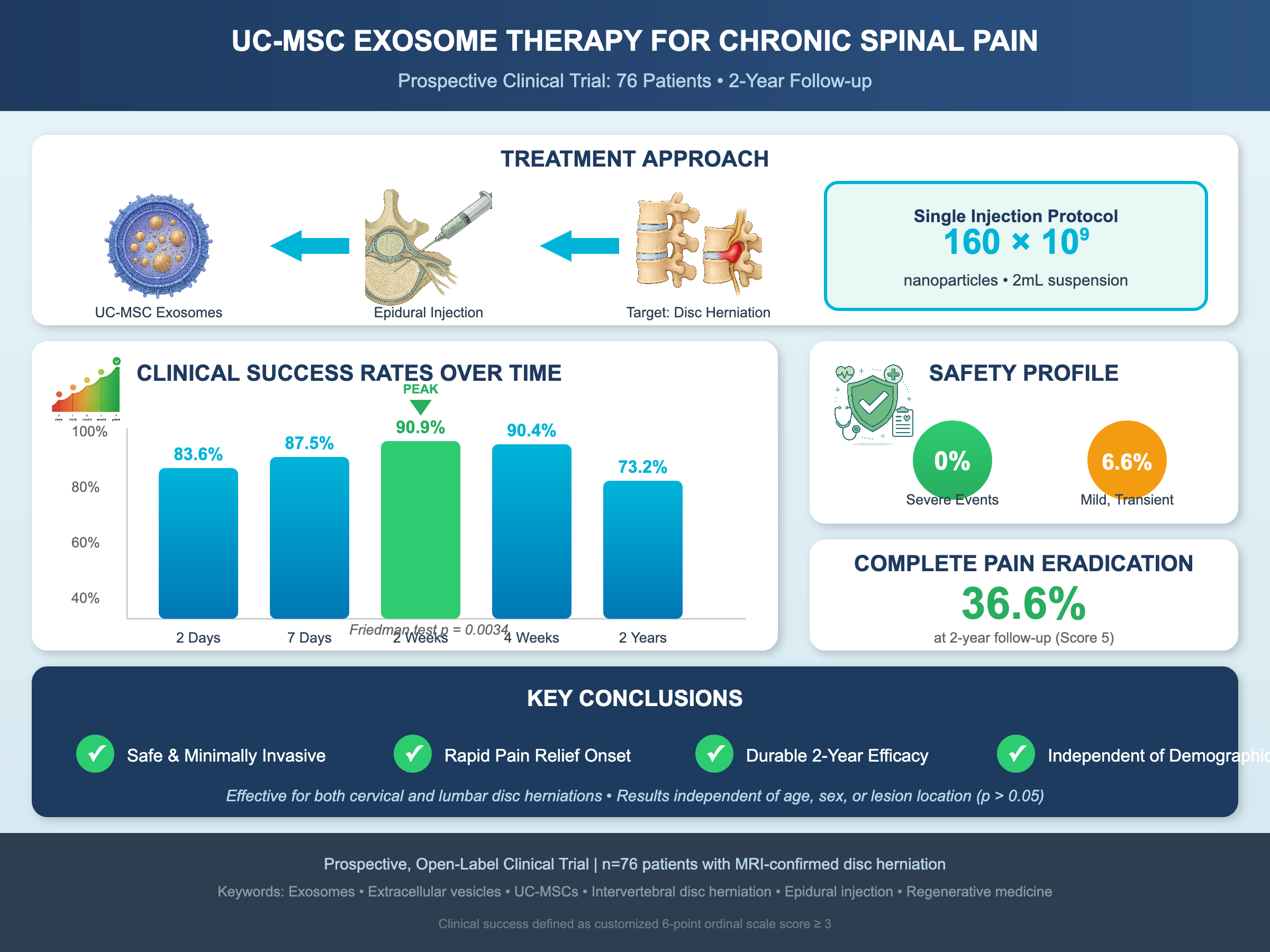
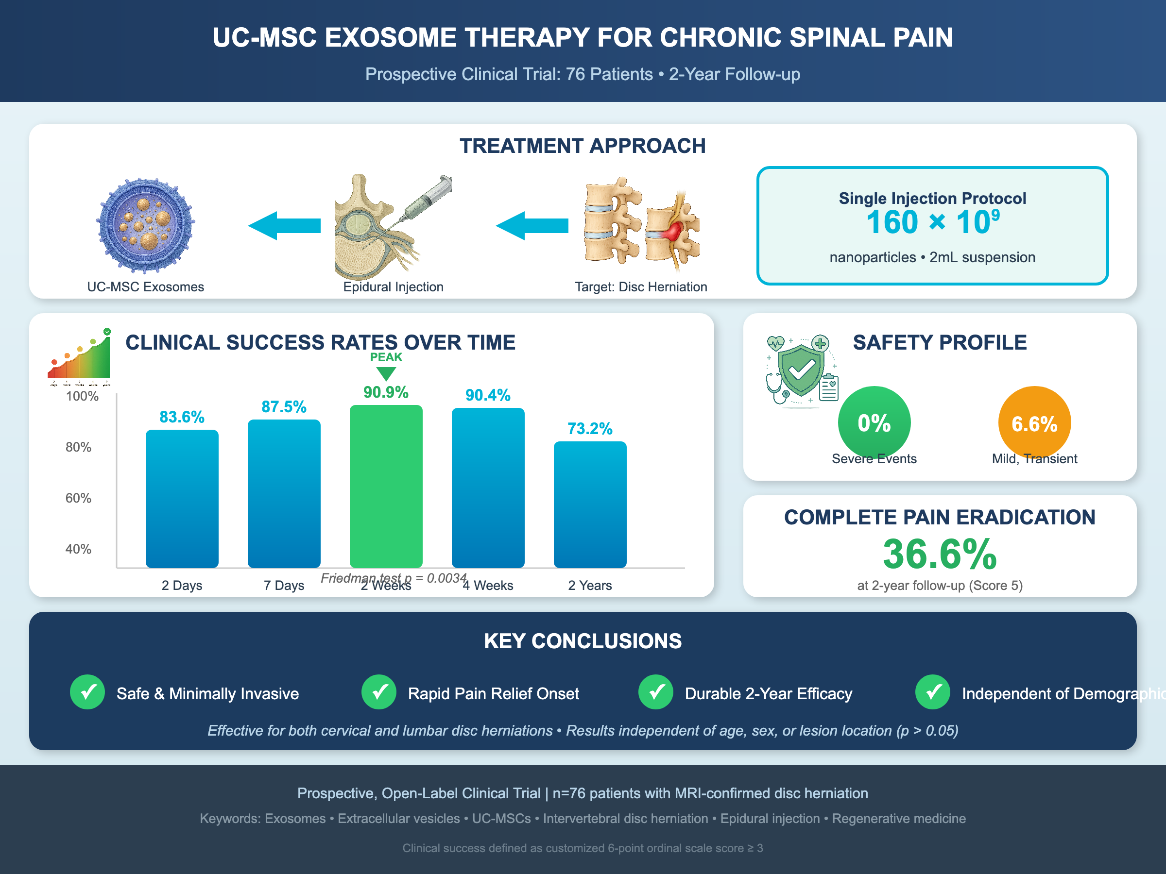
Results: No severe systemic, neurological, or infectious adverse events (0%) were observed throughout the 2-year observation period; only 6.6% of patients experienced mild, transient localized injection-site pain that resolved spontaneously within 48 hours. Clinical success rates were 83.6% at 2 days post-injection, peaking at 90.9% at 2 weeks, and remaining robust at 90.4% at 4 weeks. At the 2-year milestone, the overall success rate was maintained at 73.2%, with 36.6% of patients achieving complete pain eradication (score 5). Serial pain reduction was statistically significant over time (Friedman test, p = 0.0034) and operated independently of patient age, sex, or anatomical lesion location (p > 0.05).
Conclusion: Epidural administration of UC-MSC-Exos is a safe, minimally invasive, and effective biotherapeutic intervention. It confers rapid, sustained, and durable long-term pain relief in cervical and lumbar disc herniations, operating independently of patient demographics or anatomical variables.
Graphical abstract
INTRODUCTION
Chronic spinal pain, manifesting predominantly as intractable lower back pain with accompanying sciatica or cervical radiculopathy, constitutes a colossal global health crisis. It remains a primary contributor to years lived with disability (YLD) worldwide, imposing immense socioeconomic burdens through direct healthcare expenditures, lost workplace productivity, and profound degradation of patient quality of life1,2. The structural disruption of the intervertebral disc—specifically the herniation of the nucleus pulposus (HNP) through the annulus fibrosus—serves as the preeminent pathophysiological substrate for this chronic pain state. Within the rapidly evolving demographic landscape of the Lao People's Democratic Republic (Lao PDR), the incidence of symptomatic intervertebral disc herniation (IVDH) is ascending precipitously, intricately correlated with population aging, occupational physical stressors, and epidemiological shifts toward sedentary lifestyles.
The pathogenesis of radicular pain in IVDH is distinctly bipartite, driven by a synergistic interplay between mechanical compression and biochemical inflammation. Historically, symptomatology was attributed almost exclusively to mechanical compression of neural structures—including the spinal cord and exiting spinal nerve roots—by extruded disc tissue. However, contemporary molecular neurobiology has elucidated a crucial biochemical paradigm termed "chemical radiculitis"3. The nucleus pulposus is an immunologically privileged tissue, physiologically isolated from systemic immune surveillance by the intact annulus fibrosus since early embryonic development. Upon herniation into the highly vascularized epidural space, exposed nucleus pulposus material is recognized as a non-self autoantigen. This event triggers a virulent neuroinflammatory cascade characterized by rapid macrophage infiltration and microglial activation, culminating in a localized "cytokine storm" predominantly mediated by tumor necrosis factor-alpha (TNF-α), interleukin-1 beta (IL-1β), and interleukin-6 (IL-6)4,5. This inflammatory microenvironment induces severe perineural edema, demyelination, and nociceptor sensitization, generating neuropathic pain that persists even when overt mechanical compression is minimal.
Consequently, effective therapeutic strategies must target this inflammatory milieu alongside mechanical derangements. Conventional pharmacological management, primarily utilizing non-steroidal anti-inflammatory drugs (NSAIDs) and neuropathic pain stabilizers, frequently yields sub-optimal efficacy for chronic radicular pain and is associated with substantial gastrointestinal, renal, and cardiovascular morbidities during prolonged administration6.
When conservative measures fail, interventional procedures such as epidural steroid injections (ESIs) represent the conventional standard of care. Although ESIs deliver potent localized anti-inflammatory effects via phospholipase A inhibition, their therapeutic duration is notoriously brief. Furthermore, repetitive corticosteroid exposure carries substantial risks, including accelerated local cartilage degradation, suppression of endogenous collagen synthesis, systemic immunosuppression, osteoporosis, epidural lipomatosis, and devastating neurological sequelae secondary to particulate arterial embolization7,8. Conversely, surgical decompression effectively resolves structural mechanical impingement but carries intraoperative risks, requires prolonged convalescence, and introduces the risk of adjacent segment disease and failed back surgery syndrome (FBSS)—a state of persistent postoperative pain9.
In the vanguard of orthobiologics, regenerative medicine has aggressively investigated the therapeutic utility of mesenchymal stem cells (MSCs). Exhibiting multipotent differentiation and potent immunomodulatory capacity, live MSC transplantation has demonstrated promise in preclinical models. However, clinical translation faces formidable physiological and logistical hurdles. The epidural space represents a harsh microenvironment that severely compromises the viability of transplanted cells. Furthermore, direct cell therapy carries non-trivial risks, including ectopic osteogenesis (uncontrolled bone formation), microvascular occlusion, immune rejection, and potential tumorigenesis10.
A major paradigm shift occurred with the discovery that the therapeutic efficacy of MSCs is mediated primarily through paracrine secretion rather than direct cellular engraftment, driven largely by extracellular vesicles known as exosomes11. Exosomes are nanometer-sized (30–150 nm) lipid-bilayer vesicles originating from the endosomal compartment. They encapsulate a rich payload of bioactive molecules, including functional proteins, lipids, messenger RNAs (mRNAs), and regulatory microRNAs (miRNAs)12.
As intercellular signaling vectors, MSC-derived exosomes replicate the immunomodulatory and regenerative footprint of parent cells while avoiding the hazards of live-cell transplantation. They reprogram pro-inflammatory M1 macrophages toward the tissue-repairing M2 phenotype, suppressing neuroinflammation13,14. Concurrently, MSC-exosomes stimulate native annulus fibrosus cell proliferation, inhibit apoptosis, and upregulate extracellular matrix (ECM) synthesis, fostering tissue repair15. Utilizing exosomes derived from human umbilical cord mesenchymal stem cells (UC-MSC-Exos) offers key manufacturing advantages. As cell-free entities lacking living nuclei and major histocompatibility complex (MHC) class II antigens, UC-MSC-Exos possess minimal immunogenicity, zero risk of malignant transformation, and can be standardized as an "off-the-shelf" biological product16.
Despite compelling preclinical evidence, human clinical data evaluating epidural exosome therapy for spinal pathologies remain sparse, with no documented studies evaluating UC-MSC-Exos for discogenic pain in Lao PDR.
To address this gap, we conducted a prospective clinical trial evaluating single-dose epidural UC-MSC-Exo administration in patients with NSAID-refractory chronic pain secondary to cervical or lumbar disc herniation over a 2-year follow-up period.
MATERIALS AND METHODS
Study Design, Setting, and Ethical Considerations
This study was conducted as a prospective, open-label, single-arm clinical trial between 2023 and 2025 at NewLife Hospital, Vientiane, Lao People's Democratic Republic. The clinical protocol complied with the ethical guidelines of the Declaration of Helsinki. Ethical approval and operational clearance were granted by the Institutional Review Board (IRB) of NewLife Hospital and relevant national health regulatory authorities of Lao PDR. Written informed consent was obtained from all participants prior to enrollment after a full explanation of the procedure, potential risks, and follow-up requirements. Clinical trial registration number: Not applicable.
Patient Selection and Eligibility Criteria
A consecutive cohort of 76 patients was prospectively enrolled based on defined clinical and radiological criteria.
Inclusion Criteria:
-
Confirmed diagnosis of cervical or lumbar intervertebral disc herniation, documented by high-resolution magnetic resonance imaging (MRI) within the preceding 3 months.
-
Clinical symptoms anatomically corresponding to the MRI-identified disc lesion (e.g., chronic low back pain with radiculopathy/sciatica, or neck pain with cervicobrachial radiculopathy).
-
Symptom duration exceeding 3 months (chronic spinal pain).
-
Baseline pain severity categorized as moderate to severe, defined as a score ≥ 5 on the Wong–Baker FACES Pain Rating Scale.
-
Inadequate response or clinical failure following conservative pharmacological therapy, including an adequate trial of NSAIDs.
-
Willingness to receive a single epidural injection and adhere to the scheduled longitudinal follow-up protocol.
Exclusion Criteria:
-
Administration of additional interventional spinal injections during the study observation period.
-
Severe or rapidly progressive neurological deficits (e.g., cauda equina syndrome, severe motor weakness) requiring immediate surgical decompression.
-
Active systemic infection, localized infection at the injection site, or active malignancy.
-
Coagulopathy or severe bleeding disorders.
-
Inability or unreachability for remote telephonic follow-up assessments.
Biomanufacturing and Characterization of UC-MSC-Exosomes
The investigational UC-MSC-Exo product was manufactured using a closed-loop bioprocessing protocol under Good Manufacturing Practice (GMP) conditions.
Step 1: Primary Cell Cultivation and Conditioned Media Harvest
Human umbilical cord mesenchymal stem cells (UC-MSCs) were expanded using the MSCCult platform developed at the Stem Cell Institute (SCI), University of Science, Vietnam National University, Ho Chi Minh City, Vietnam. Cells were expanded to passage 7 (P7). Upon reaching 70% confluence, the culture medium was aspirated, washed, and replaced with serum-free Dulbecco's Modified Eagle Medium (DMEM) at a volume of 1 mL per 10 cells. Cells were incubated for 24 hours to generate exosome-rich conditioned media (CM). All cell processing was performed in an ISO Class 5 cleanroom environment.
Step 2: Exosome Isolation and Nanoscale Concentration
Harvested CM was processed using an automated ultrasonic nanofiltration system. This system combines negative pressure oscillation with ultrasound waves to concentrate exosomes 80- to 100-fold while eliminating >99% of soluble host proteins and cell debris.
Step 3: Quality Control, Characterization, and Standardization
Prior to clinical release, concentrated exosome preparations underwent biophysical and safety testing:
-
Particle Size and Concentration: Evaluated by nanoparticle tracking analysis (NTA; Nanosight).
-
Surface Marker Profiling: Flow cytometry confirmed expression of canonical tetraspanin markers (CD9, CD63, and CD81).
-
Biosafety Testing: Tested for Mycoplasma, bacterial contamination (aerobic/anaerobic), and endotoxin levels using the Limulus Amebocyte Lysate (LAL) assay. Pyrogenicity was evaluated using the Monocyte Activation Test (MAT).
Following quality control release, the final formulation was standardized to a working concentration of 80 × 10 nanoparticles/mL.
Clinical Epidural Intervention Protocol
Interventions were performed under sterile conditions in an operating theater. Guided by procedural protocols established by the Ministry of Health of Vietnam (2014), an interventional specialist placed an epidural needle into the target epidural space using the loss-of-resistance technique. Following negative aspiration for cerebrospinal fluid and blood, a single 2 mL dose of standardized UC-MSC-Exo suspension (total payload: 160 × 10 nanoparticles) was slowly injected adjacent to the herniated disc segment.
Clinical Evaluation Parameters and Outcome Measures
Clinical evaluations were conducted via structured telephonic interviews by clinical coordinators at predefined intervals: +2 days, +7 days, +2 weeks, +4 weeks, and +2 years post-injection.
Efficacy Assessment (Pain and Function):
Assessed using a customized 6-point ordinal grading scale:
-
Score 0: Severe pain, no improvement.
-
Score 1: Severe pain, negligible improvement.
-
Score 2: Severe pain, noticeable relief.
-
Score 3: Moderate pain, significant improvement (Good Response).
-
Score 4: Mild pain, no interference with daily activities.
-
Score 5: Complete or near-complete pain relief (Excellent Response).
Treatment success ("Clinical Success") was defined as achieving a score ≥ 3.
Safety Assessment:
Adverse events (AEs) were monitored, including injection-site pain, infection, meningeal irritation, CSF leakage, and systemic allergic or anaphylactic reactions.
Statistical Analysis
Statistical analysis was performed using GraphPad Prism software. Ordinal pain scores were analyzed using non-parametric methods. Changes in pain scores across time points were evaluated using the Friedman test, followed by post-hoc pairwise comparisons using the Wilcoxon signed-rank test with Bonferroni correction. Subgroup comparisons (anatomical level, age, sex) were performed using the Kruskal–Wallis test and Mann–Whitney U test. A two-tailed p-value < 0.05 was considered statistically significant.
RESULTS
In Vitro Characterization of UC-MSC-Exosomes
NTA characterization demonstrated a uniform nanoparticle population with a mean size of 113.4 ± 1.4 nm, a mode of 98.7 ± 2.8 nm, and a standard deviation of 40.0 ± 3.1 nm. Stock concentration prior to final formulation was (8.52 ± 0.37) × 10 particles/mL (Figure 1A).
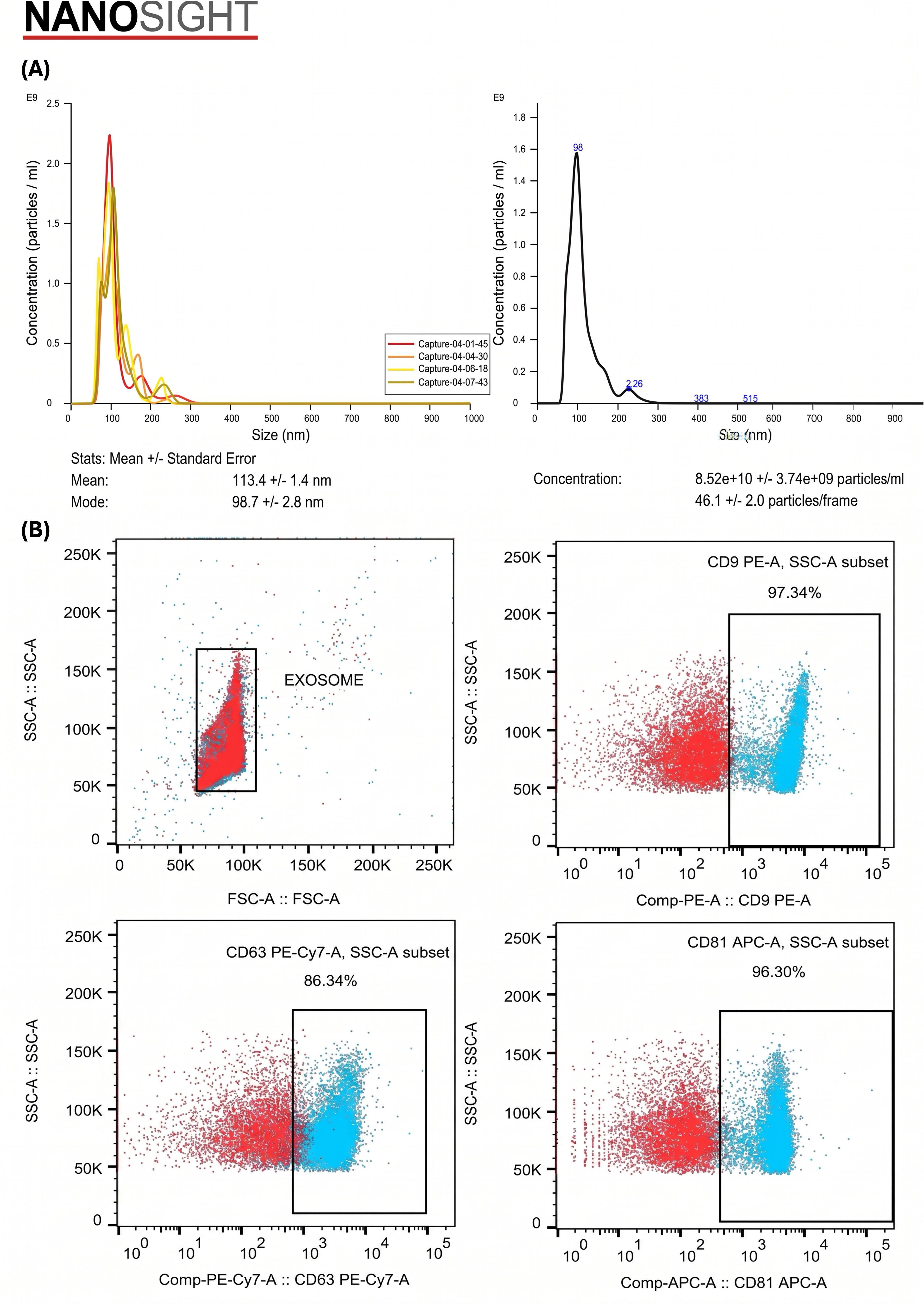
Biophysical characterization and marker expression of clinical-grade umbilical cord mesenchymal stem cell-derived exosomes (UC-MSC-Exos).(A) Particle size distribution and absolute concentration measured by Nanosight nanoparticle tracking analysis (NTA), demonstrating a sharp, homogeneous vesicular profile with a mean particle diameter of 113.4 ± 1.4 nm and mode of 98.7 ± 2.8 nm (concentration: 8.52 × 10¹⁰ ± 3.74 × 10⁹ particles/mL). (B) Flow cytometry phenotyping showing robust positive expression for canonical exosomal tetraspanins CD9 (97.34%), CD63 (86.34%), and CD81 (96.30%). Gates were defined using isotype controls. Data represent quality control validation prior to clinical epidural administration.
Flow cytometry confirmed high expression of tetraspanin markers: CD9 (97.34%), CD63 (86.34%), and CD81 (96.30%) (Figure 1B). Biosafety assays confirmed negative results for Mycoplasma, bacterial contamination, endotoxins, and pyrogenic activity.
Patient Demographics and Baseline Characteristics
The final study cohort comprised 76 enrolled patients (Figure 2). Demographics showed an even sex distribution (37 males [48.7%], 39 females [51.3%]) (Table 1). Age distribution was as follows: < 40 years (22.4%), 40–59 years (39.5%), and ≥ 60 years (38.2%).
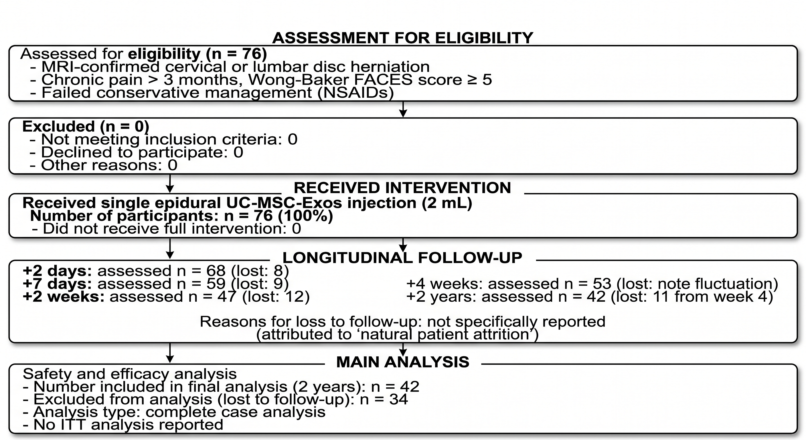
Flow diagram of participant eligibility, enrollment, epidural intervention, and 2-year longitudinal follow-up. Schematic flowchart detailing patient screening (N = 76), single-dose epidural UC-MSC-Exo administration (2 mL; 160 × 109 total nanoparticles), and retained patient numbers across five temporal follow-up milestones (+2 days, +7 days, +2 weeks, +4 weeks, and +2 years). Natural attrition rates and final complete-case analysis numbers (n = 42 at 2 years) are specified.
Baseline anthropometric, demographic, and clinical characteristics of the study cohort (N = 76) and subgroup efficacy comparisons.
| Parameter | Value (N = 76) | Percentage (%) | P-value (Efficacy difference) |
|---|---|---|---|
| Sex | |||
| Male | 37 | 48.7% | |
| Female | 39 | 51.3% | |
| Age Group (Years) | |||
| < 40 | 17 | 22.4% | |
| 40–59 | 30 | 39.5% | |
| ≥ 60 | 29 | 38.2% | |
| Baseline Clinical Status | |||
| Pain Chronicity | > 3 months | 100% | |
| Pain Severity (Wong–Baker FACES) | ≥ 5 | 100% | |
Topographically, herniations were most frequent in the lower lumbar spine (Figure 3): L4–L5 (48.8%) and L5–S1 (23.8%). Cervical herniations were predominantly at C5–C6 (16.7%), C4–C5 (3.6%), and C6–C7 (1.2%). Other locations included L3–L4 (4.8%) and T12–L1 (1.2%).
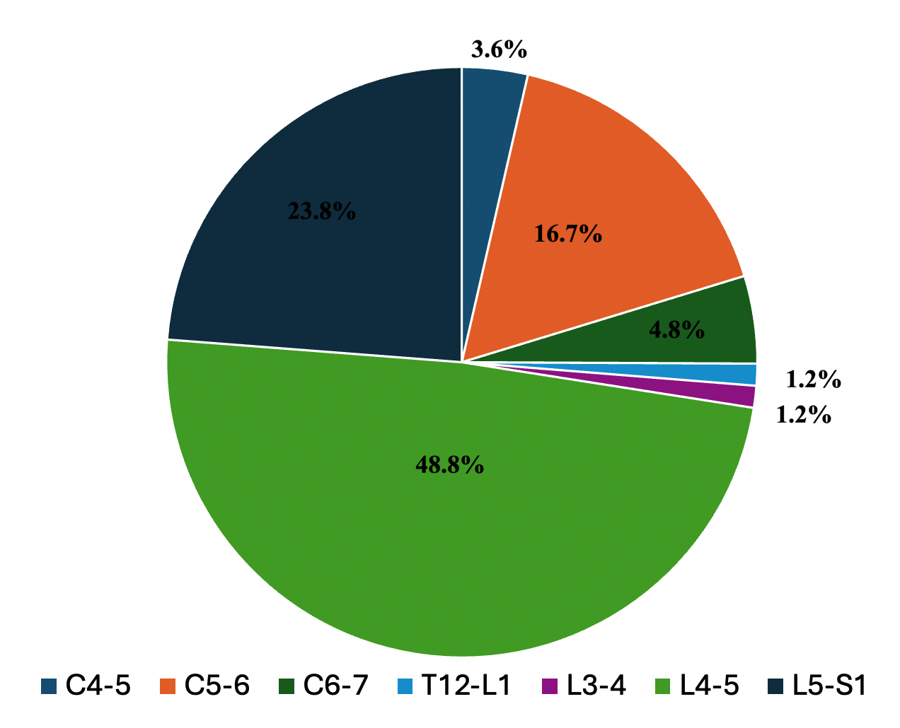
Anatomical distribution of intervertebral disc herniation segments across the study cohort (N = 76). Pie chart depicting the proportion of targeted disc herniation levels in the cervical, thoracolumbar, and lumbar spinal regions. Lower lumbar segments accounted for the majority of cases (L4–L5: 48.8%, L5–S1: 23.8%), followed by cervical segments (C5–C6: 16.7%). Non-parametric subgroup analysis (Kruskal–Wallis test) confirmed that analgesic efficacy was independent of anatomical lesion level (
Safety Profile Evaluation
No severe systemic, neurological, or infectious complications (0%) occurred over the 2-year follow-up period (no meningitis, epidural abscess, CSF leakage, or anaphylaxis).
The only observed adverse event was mild, transient injection-site pain in 5 patients (6.6%), which resolved spontaneously within 24 to 48 hours without intervention.
Longitudinal Efficacy and Pain Trajectory
Assessed patient numbers across follow-up intervals were: n = 68 at 2 days, n = 59 at 7 days, n = 47 at 2 weeks, n = 53 at 4 weeks, and n = 42 at 2 years (Figure 2). Pain score progression demonstrated distinct clinical phases (Figure 4).
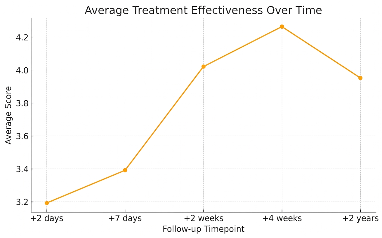
Longitudinal clinical efficacy and temporal trajectory of pain alleviation over 2 years post-epidural injection. Line graph illustrating changes in mean pain improvement score (evaluated on a 0–5 ordinal scale) across predefined follow-up time points (+2 days, +7 days, +2 weeks, +4 weeks, and +2 years). Peak average improvement occurred at 4 weeks (mean score = 4.25), reflecting acute neuroinflammatory suppression, followed by sustained long-term pain relief up to 2 years (mean score = 3.95). Overall variance across time points was statistically significant (Friedman test,
Acute Phase (0 to 2 Weeks):
At 2 days post-injection, mean improvement score was 3.2, with an 83.6% clinical success rate (score ≥ 3) and 3.0% reporting complete pain relief (score 5). By 2 weeks, mean score increased to 4.0, success rate reached 90.9%, and complete pain relief increased to 27.3%.
Medium-Term Phase (4 Weeks):
At 4 weeks, mean score reached 4.25, with a 90.4% success rate and 19.2% reporting complete pain relief.
Long-Term Phase (2 Years):
At 2 years (n = 42), the overall success rate was 73.2% (mean score = 3.95). Notably, the proportion of patients with complete pain relief (score 5) reached its highest level at 36.6%. Treatment failure (scores 0–1) occurred in a minority of patients due to progressive degenerative changes.
The Friedman test indicated significant overall variance across time points (χ = 15.72, p = 0.0034). Post-hoc analyses showed scores at 2 weeks, 4 weeks, and 2 years were significantly higher than at 2 days (p < 0.05), with no significant degradation between 2 weeks, 4 weeks, and 2 years.
Subgroup Analyses
-
Anatomical Location: Kruskal–Wallis test showed no significant differences in efficacy among L4–L5, L5–S1, and C5–C6 herniations (
p > 0.05). -
Age and Sex: Pain relief was comparable across age groups (< 40, 40–59, ≥ 60 years) and between male and female patients (
p > 0.05). Spearman correlation confirmed no inverse relationship between age and pain improvement.
DISCUSSION
This prospective trial evaluates epidural UC-MSC-Exo administration for chronic discogenic pain in Lao PDR, featuring a 2-year follow-up. The results indicate that this cell-free biological therapy is safe and effective.
The absence of severe complications highlights advantages of exosomes over live stem cell therapy. Being non-replicating, cell-free vesicles, exosomes avoid risks of ectopic ossification, microvascular embolization, and tumorigenesis10. Lacking MHC class II antigens, UC-MSC-Exos show minimal immunogenicity, reducing rejection risks16. Injection-site discomfort (6.6%) was transient and mechanical rather than immunogenic.
Clinical kinetics showed a biphasic response. Early pain reduction (90.9% success at week 2) reflects acute anti-inflammatory effects rather than structural repair. Herniated disc material causes chemical radiculitis by recruiting M1 macrophages4. MSC-exosomes contain microRNAs (e.g., miR-146a, let-7b) that inhibit NF-κB signaling and promote M1-to-M2 macrophage polarization13,14, reducing perineural edema and hyperalgesia.
The long-term phase showed sustained relief, with 36.6% achieving complete pain resolution at 2 years. Exosomes deliver trophic factors (e.g., TGF-β, VEGF) that may inhibit apoptosis, promote matrix synthesis, and support tissue remodeling15. Initial exosome administration may initiate self-sustaining tissue repair in responsive patients. Conversely, late treatment failures likely reflect mechanical limits of biotherapeutics in severe structural degeneration.
Compared to epidural steroids, which carry catabolic risks and short-lived effects7, UC-MSC-Exos provide immunomodulatory and regenerative cues. Unlike autologous PRP, which exhibits donor variability12, UC-MSC-Exos offer a standardized off-the-shelf preparation.
Analgesic efficacy was consistent across cervical and lumbar regions and independent of patient age, indicating broad applicability even in older individuals with reduced endogenous repair capacity.
Limitations: The open-label, single-arm design cannot exclude placebo effects, though sustained relief at 2 years reduces this likelihood. Telephonic follow-ups lack physical examination detail, and the absence of follow-up MRI limits radiological correlation. Future randomized controlled trials with imaging endpoints are recommended.
CONCLUSION
Epidural administration of UC-MSC-Exos is a safe, minimally invasive, and effective intervention for chronic radicular pain secondary to cervical and lumbar disc herniation. This cell-free approach avoids live-cell therapy risks, providing rapid neuroinflammatory suppression followed by long-term clinical relief. Therapeutic efficacy was consistent across patient age, sex, and lesion location. Randomized controlled trials are warranted to further validate these findings.
DECLARATIONS
Abbreviations
AE: Adverse event; CM: Conditioned media; CSF: Cerebrospinal fluid; DMEM: Dulbecco's modified Eagle medium; ECM: Extracellular matrix; ESI: Epidural steroid injection; FBSS: Failed back surgery syndrome; GMP: Good manufacturing practice; HNP: Herniation of the nucleus pulposus; IL: Interleukin; IVDH: Intervertebral disc herniation; LAL: Limulus amebocyte lysate; MAT: Monocyte activation test; MHC: Major histocompatibility complex; MRI: Magnetic resonance imaging; MSC: Mesenchymal stem cell; NSAIDs: Non-steroidal anti-inflammatory drugs; NTA: Nanoparticle tracking analysis; PRP: Platelet-rich plasma; TNF-α: Tumor necrosis factor-alpha; TRPS: Tunable resistive pulse sensing; UC-MSC-Exos: Umbilical cord-derived mesenchymal stem cell exosomes; YLD: Years lived with disability.
Acknowledgments
None.
Author’s contributions
N.C.P. served as the principal clinical investigator, taking the primary role in clinical interventions, patient assessments, data analysis, and drafting the manuscript. N.B.V. provided essential biological materials and technological support (UC-MSC-Exos and MSCCult platform), conducted laboratory validation for exosomes, performed data analysis, supervised the methodology, and finalized the manuscript. All authors have read and approved the final manuscript.
Funding
None.
Availability of data and materials
The datasets used and/or analyzed during the current study are available from the corresponding author on reasonable request.
Ethics approval and consent to participate
This study was approved by the Institutional Review Board (IRB) of NewLife Hospital and the regulatory health authorities of Lao PDR. Written informed consent was obtained from all individual participants included in the study.
Consent for publication
Not applicable.
Declaration of generative AI and AI-assisted technologies in the writing process
The authors declare that they used generative AI and AI-assisted technologies (Gemini) during the writing process prior to submission to improve language fluency and readability. All content was reviewed and finalized by the authors, who take full responsibility for the integrity of the published work.
Competing interests
The authors declare that they have no competing interests.