


In Vitro Antitumor Effects of the Hydroalcoholic Extract of Onopordum leptolepis on SW480 Colorectal Cancer Cells
- Department of Operating Room, Nahavand School of Allied Medical Sciences, Hamadan University of Medical Sciences, Hamadan, Iran
- Department of Anatomical Sciences, School of Medicine, Kermanshah University of Medical Sciences, Kermanshah, Iran
- Student Research Committee of Kermanshah University of Medical Sciences, Kermanshah, Iran
Abstract
Introduction: Colorectal carcinoma (CRC) is among the most prevalent and lethal malignancies worldwide. Because conventional chemotherapy is frequently limited by severe systemic adverse effects and the emergence of chemoresistance, alternative and complementary therapeutic strategies are under active investigation. This study investigated the in vitro anticancer activity of a 70% ethanol hydroalcoholic extract of Onopordum leptolepis against SW480 colorectal cancer cells and its association with modulation of the Notch1/Jagged1/c-Myc signaling pathway.
Methods: A 70% ethanol hydroalcoholic extract of O. leptolepis was prepared by maceration. Human SW480 CRC cells and non-tumorigenic L929 mouse fibroblasts were cultured. Cell viability following exposure to increasing extract concentrations (12.5–800 µg/mL) was evaluated using the MTT assay, and half-maximal inhibitory concentrations (IC50) were estimated via nonlinear regression. Apoptosis was quantified through diphenylamine-based DNA fragmentation analysis. Relative mRNA expression levels of Notch1, Jagged1, and c-Myc were determined by quantitative reverse transcription PCR (qRT-PCR), and cellular protein levels were measured by ELISA. Cell motility and invasiveness were evaluated using scratch wound-healing and Matrigel-coated Transwell assays, respectively.
Results: The hydroalcoholic extract of O. leptolepis exerted concentration-dependent cytotoxicity against SW480 cells with an IC50 of 290.11 µg/mL (95% CI: 259.8–324.0 µg/mL), whereas L929 fibroblasts exhibited marked resistance (viability > 75% at 800 µg/mL; IC50 > 800 µg/mL). Treatment of SW480 cells for 24 h caused a significant, dose-dependent decrease in viability, declining from 88–92% at 12.5–50 µg/mL (p < 0.05) to 55–70% at 100–200 µg/mL and 32% at 800 µg/mL (all p < 0.001). Apoptotic DNA fragmentation increased progressively from 8–12% at 12.5–25 µg/mL (p < 0.05) to 30–42% at 100–200 µg/mL, reaching nearly 60% at 800 µg/mL (p < 0.001). Extract treatment significantly downregulated Notch1, Jagged1, and c-Myc at both the mRNA and cellular protein levels (all p < 0.001). Furthermore, cell migration was markedly suppressed, with scratch wound closure reduced from 45–50% in controls to 20–25% in treated cells (p < 0.001), and Transwell Matrigel invasion was significantly inhibited (p < 0.001).
Conclusion: The 70% ethanol hydroalcoholic extract of O. leptolepis demonstrates significant in vitro antiproliferative, pro-apoptotic, antimigratory, and anti-invasive properties in SW480 colorectal cancer cells, mediated in part through suppression of the Notch1/Jagged1/c-Myc signaling pathway. These findings highlight O. leptolepis as a promising botanical candidate for further phytochemical isolation and translational evaluation in colorectal cancer management.
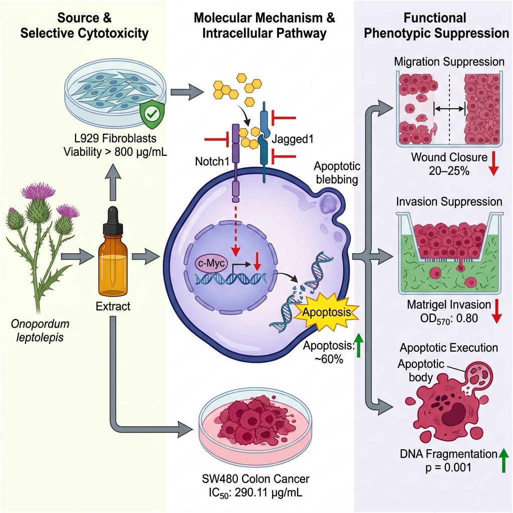
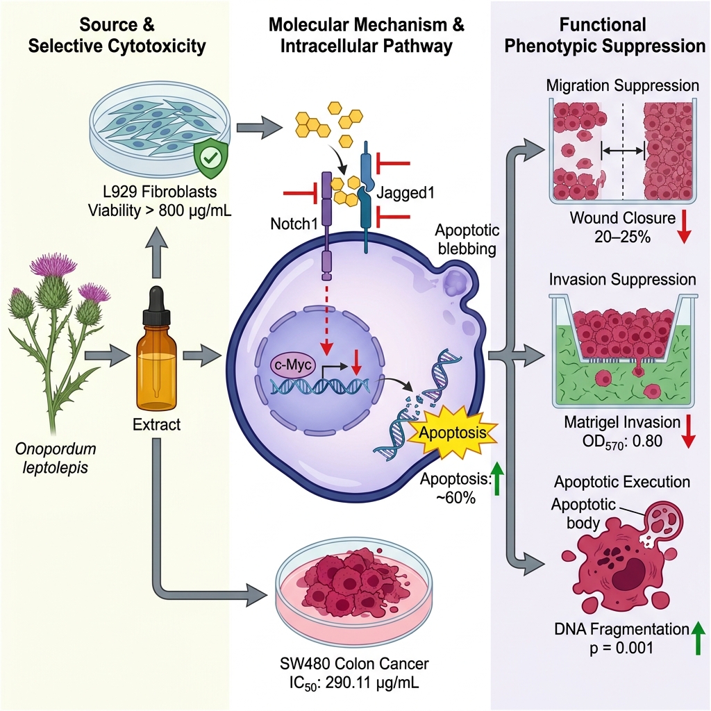
Graphical abstract
Introduction
Colorectal cancer (CRC) remains one of the leading causes of cancer-related morbidity and mortality worldwide. According to the GLOBOCAN 2022 estimates published by the International Agency for Research on Cancer (IARC), more than 1.9 million new CRC cases and over 900,000 deaths occurred globally in 2022, establishing CRC as the third most commonly diagnosed malignancy and the second leading cause of cancer mortality worldwide1. In Iran, national cancer registry and epidemiological data indicate approximately 131,000–140,000 new cancer diagnoses and more than 70,000 cancer-related deaths annually2. Among these malignancies, CRC poses a formidable public health challenge and is recognized as one of the most prevalent tumors of the gastrointestinal tract3. The high invasive and metastatic propensity of CRC represents a major determinant of adverse clinical prognosis and remains a primary cause of treatment failure despite advancements in diagnostic screening and therapeutic interventions4.
Although systemic chemotherapy remains a cornerstone of CRC management, its clinical utility is frequently hindered by severe off-target cytotoxicities and the eventual emergence of multidrug resistance during prolonged therapy. These limitations underscore the urgent need to identify novel therapeutic candidates with enhanced antitumor efficacy and favorable safety profiles5. In recent years, numerous plant-derived bioactive compounds and traditional herbal formulations have exhibited potent anticancer activities through the induction of apoptosis, suppression of tumor cell proliferation, inhibition of migration and invasion, and modulation of key oncogenic signaling cascades, including the Notch, PI3K/Akt, and NF-κB pathways. For example, flavonoids isolated from Ageratum conyzoides6 and the traditional formulation Miao-Yi-Ai-Tang7 have demonstrated marked potential in halting cervical and lung cancer progression, respectively. Onopordum leptolepis, commonly known in Persian as “Kharpanbeh”, is a perennial species belonging to the Asteraceae family that thrives predominantly in semi-arid environments8. Morphologically, it is distinguished by pink inflorescences, spiny aerial structures, compartmentalized receptacles, and glabrous, sub-quadrangular achenes. Historically, this species has been utilized in traditional medicine for various therapeutic applications. Owing to its rich composition of flavonoids and phenolic derivatives, O. leptolepis exhibits notable pharmacological activities, including potent antioxidant and antiproliferative properties, and has been investigated as a protective constituent in dermatological and pharmaceutical preparations9.
The Notch signaling pathway is an evolutionarily conserved molecular cascade critically implicated in regulating embryonic development, tissue homeostasis, and diverse tumorigenic processes, including cellular proliferation, differentiation, survival, angiogenesis, migration, and invasion. Canonical Notch signaling is initiated through interactions between transmembrane Notch receptors (Notch1–4) and membrane-bound ligands (Jagged1/2 and Delta-like 1/3/4), triggering sequential proteolytic cleavages that release the active Notch intracellular domain (NICD) to translocate into the nucleus and drive target gene transcription10. Among these pathway constituents, Notch1, its ligand Jagged1, and the downstream proto-oncogenic effector c-Myc are critically involved in promoting CRC cell proliferation, inhibiting apoptosis, and driving metastatic dissemination11. Accumulating clinical and experimental evidence indicates that aberrant overexpression of Notch1 and Jagged1 correlates strongly with disease recurrence, chemoresistance, and metastasis in CRC, suggesting that deregulated Notch signaling serves as an essential driver of colorectal tumorigenesis.
Although various phytochemicals have been reported to inhibit Notch signaling in colorectal malignancies, the biological activity and molecular targets of O. leptolepis remain largely unexplored. To our knowledge, no previous investigation has characterized the antitumor effects of the 70% ethanol hydroalcoholic extract of O. leptolepis in CRC cells or evaluated its potential association with the Notch1/Jagged1/c-Myc signaling pathway. Therefore, the present study aimed to evaluate the in vitro antiproliferative, pro-apoptotic, antimigratory, and anti-invasive properties of O. leptolepis extract in SW480 human colorectal cancer cells and to determine whether these phenotypic anticancer actions are associated with suppression of the Notch signaling cascade.
Methods
Preparation of the 70% Ethanol Hydroalcoholic Extract of Onopordum leptolepis
Plant specimens of Onopordum leptolepis were collected from the mountainous regions of Kermanshah, Iran (GPS coordinates: 34° 18' 51.01" N, 47° 03' 54.00" E) during the optimal flowering season. Botanical authentication was confirmed by an expert plant taxonomist, and a voucher specimen was deposited in the institutional herbarium (Voucher No. KU-OV-2020-001). The aerial parts (leaves and flowering stems) were cleaned and shade-dried at ambient room temperature to prevent the thermal degradation of thermolabile phytoconstituents. The dried material (40 g) was pulverized into a fine powder using an electric grinder to optimize surface area contact and enhance solvent penetration during extraction. Hydroalcoholic extraction was performed using the maceration technique with 70% ethanol (v/v), a solvent system established for efficiently extracting a broad spectrum of polar and semi-polar bioactive phytoconstituents. The pulverized plant material was immersed in 300 mL of 70% ethanol and agitated on an orbital shaker at moderate speed for 72 h at room temperature. The mixture was filtered through Whatman No. 1 filter paper to remove insoluble particulate residues. The filtrate was concentrated under a chemical fume hood with gentle airflow at room temperature and subsequently dried in a temperature-controlled oven at 45 °C for 24 h to ensure complete removal of residual ethanol. The final dried crude extract was stored in airtight, light-protected containers at −20 °C until experimental use. Stock solutions were prepared by dissolving the dried extract in dimethyl sulfoxide (DMSO) and subsequently diluted with culture medium to achieve designated working concentrations. The final DMSO concentration in all treatment wells was maintained at ≤ 0.1% (v/v), a level confirmed to exert negligible cytotoxicity. Vehicle control wells received an equivalent concentration of DMSO (0.1%) without plant extract12.
Cell Culture Conditions
Human colorectal carcinoma cells (SW480) and non-tumorigenic mouse subcutaneous fibroblasts (NCTC clone 929 / L929) were obtained from established cell repositories and cultured under standard sterile conditions in 25-cm² tissue culture flasks. SW480 cells were maintained in RPMI-1640 medium supplemented with 10% heat-inactivated fetal bovine serum (FBS) and 1% penicillin–streptomycin (100 U/mL penicillin, 100 µg/mL streptomycin). Experiments were conducted using cells between passages 4 and 10. L929 fibroblasts were cultured in Dulbecco’s Modified Eagle Medium (DMEM) enriched with 10% FBS and 1% penicillin–streptomycin. Both cell lines were maintained at 37 °C in a humidified atmosphere containing 5% CO, with growth media replenished every 48–72 h. Cell morphology and confluence were routinely monitored using an inverted phase-contrast microscope, and cells were subcultured using 0.25% trypsin–EDTA upon reaching 80–90% confluence13.
Cell Viability Assessment (MTT Assay)
Cell viability was evaluated using the 3-(4,5-dimethylthiazol-2-yl)-2,5-diphenyltetrazolium bromide (MTT) colorimetric assay. Briefly, SW480 cells and L929 fibroblasts were seeded into 96-well plates at a density of 1.5 × 10 cells/well and incubated overnight to facilitate attachment and equilibration. Cells were subsequently treated with increasing concentrations of the 70% ethanol hydroalcoholic extract (12.5, 25, 50, 100, 200, 400, and 800 µg/mL) or vehicle control (0.1% DMSO) for 24 h. The concentration range was selected to encompass non-cytotoxic, moderately cytotoxic, and high-dose cytotoxic thresholds for comprehensive dose-response modeling. Following 24 h of incubation, 30 µL of MTT solution (5 mg/mL in PBS) was added to each well, and plates were incubated for an additional 3 h at 37 °C in the dark to facilitate the reduction of MTT into insoluble formazan crystals by active mitochondrial dehydrogenases. The supernatant was carefully aspirated, and 100 µL of DMSO was added to each well to solubilize the formazan precipitates. Plates were gently agitated on a microplate shaker for 20 min at room temperature to ensure complete dissolution, and absorbance was measured at 570 nm using an ELISA microplate reader. Percentage cell viability was calculated as follows: Cell Viability (%) = (OD / OD) × 100. The extract remained visually soluble throughout the 24-h incubation period without observable precipitation14.
Determination of IC Values
Cell viability data obtained after 24 h of extract exposure were fitted to a four-parameter logistic (4PL) nonlinear regression model using GraphPad Prism (version 8.0) and SPSS (version 16.0). Concentration–response curves were generated by plotting cell viability (%) against the log of extract concentration (µg/mL). The half-maximal inhibitory concentration (IC) values, corresponding 95% confidence intervals (95% CI), and goodness-of-fit coefficients (R²) were derived from the fitted regression curves.
Apoptosis Quantification (Diphenylamine-Based DNA Fragmentation Assay)
Apoptotic cell death was evaluated by quantifying low-molecular-weight DNA fragmentation using the diphenylamine (DPA) colorimetric method. Following 24 h of treatment with designated extract concentrations, approximately 5 × 10 SW480 cells were harvested, washed with cold PBS, and lysed in 1 mL of TTE lysis buffer (10 mM Tris-HCl, pH 7.4, 1 mM EDTA, 0.2% Triton X-100) to selectively release fragmented apoptotic DNA. Cell lysates were centrifuged at 20,000 × g for 10 min at 4 °C to separate fragmented DNA (supernatant, Sample A) from intact, high-molecular-weight chromatin (pellet, Sample B). The supernatant was collected into a fresh tube, while the pellet was resuspended in 1 mL of TTE buffer. Both fractions were combined with an equal volume of 25% trichloroacetic acid (TCA) and incubated overnight at 4 °C to precipitate DNA. Following centrifugation at 20,000 × g for 10 min, the resulting precipitates were hydrolyzed by adding 160 µL of 5% TCA and heating at 90 °C for 15 min. Subsequently, 320 µL of freshly prepared DPA reagent was added to each sample, followed by incubation at 37 °C for 4 h to allow color development. Optical density was measured at 600 nm using a spectrophotometer. The percentage of apoptotic DNA fragmentation was calculated using the formula: DNA Fragmentation (%) = [OD / (OD + OD)] × 10015.
Gene Expression Analysis by Quantitative Real-Time PCR (qRT-PCR)
Total cellular RNA was extracted from treated and untreated SW480 cells using TRIzol reagent according to the manufacturer’s instructions and stored at −80 °C. RNA concentration and purity were assessed using a NanoDrop spectrophotometer, with A/A absorbance ratios between 1.8 and 2.0 considered acceptable for downstream applications. Complementary DNA (cDNA) was synthesized from 1 µg of total RNA using the RevertAid First Strand cDNA Synthesis Kit (Thermo Fisher Scientific, USA; Cat. No. K1622). Quantitative real-time PCR was performed using PowerUp™ SYBR™ Green Master Mix (Applied Biosystems, USA; Cat. No. A25742) on a real-time PCR detection system. The amplification protocol consisted of an initial denaturation at 95 °C for 10 min, followed by 40 cycles of denaturation at 95 °C for 15 s, annealing at 60 °C for 30 s, and extension at 72 °C for 30 s. A terminal melting curve analysis (65–95 °C) was conducted to verify amplification specificity and confirm the absence of primer-dimer artifacts. The specific primer sequences were as follows:
-
Notch1 16: Forward 5′-CGTTCCAGCAGTCTCCGTC-3′, Reverse 5′-GTGGGCCAGTCTCAAAGG-3′ -
Jagged1 16: Forward 5′-AGTGCCTGAATGGACGGA-3′, Reverse 5′-TGGAGACTGGAAGACCGA-3′ -
c-Myc 17: Forward 5′-TCTCCATCCTATGTTGCGGTC-3′, Reverse 5′-TCCAAGTAACTCGGTCATCATCT-3′ -
GAPDH 18: Forward 5′-GTCTCCTCTGACTTCAACAGCG-3′, Reverse 5′-ACCACCCTGTTGCTGTAGCCAA-3′
GAPDH served as the internal housekeeping control. Relative mRNA expression levels were calculated using the comparative 2 method19.
Quantification of Notch1, Jagged1, and c-Myc Protein Levels by ELISA
Cellular protein levels of Notch1, Jagged1, and c-Myc were determined using commercially available, enzyme-linked immunosorbent assay (ELISA) kits according to the manufacturers’ protocols: human Notch1 ELISA kit (Invitrogen, Thermo Fisher Scientific; Cat. No. EHNOTCH1), human Jagged1 ELISA kit (R&D Systems; Cat. No. DY599), and human c-Myc ELISA kit (MyBioSource; Cat. No. MBS724950). Following 24 h of treatment, SW480 cells were gently detached using a non-enzymatic cell dissociation solution, washed twice with ice-cold PBS, and lysed in cell lysis buffer containing protease inhibitors. Protein extracts were added to pre-coated microplates and incubated with target-specific capture and biotinylated detection antibodies, followed by incubation with streptavidin-horseradish peroxidase conjugate. Chromogenic substrate solution was added, and absorbance was measured at 450 nm (with wavelength correction at 570 nm) using a microplate reader. Target protein concentrations were calculated from standard curves generated with serial dilutions of purified recombinant standards and normalized to total protein content. Untreated cells served as controls, and blank wells containing assay reagents without cell lysate were included to determine background absorbance20.
Scratch Wound-Healing Assay (Cell Migration)
Two-dimensional collective cell motility was evaluated using the scratch wound-healing assay. SW480 cells were seeded into 6-well plates at a density of 1.6 × 10 cells/mL and cultured until reaching a confluent monolayer. A uniform, linear cell-free scratch was generated across the center of each well using a sterile 1-mL pipette tip. Wells were washed gently with PBS to remove dislodged cells and debris. Cells were then incubated in DMEM supplemented with 2% FBS (to minimize confounding effects of cellular proliferation) in the presence or absence of O. leptolepis extract for 24 h at 37 °C. Wound closure was monitored by photographing identical coordinates immediately after scratch generation (0 h) and after 24 h using an inverted phase-contrast microscope equipped with a digital camera. Quantitative measurement of the denuded area was performed using automated threshold-based image segmentation in TScratch software (MathWorks Inc.). The percentage of wound closure was calculated as: Wound Closure (%) = [(Wound Area at 0 h − Wound Area at 24 h) / Wound Area at 0 h] × 10021.
Matrigel Transwell Invasion Assay
Cellular invasive capacity was evaluated using 24-well Transwell chambers containing 8-µm pore polycarbonate membrane inserts (6.5-mm diameter; Corning Inc.). The upper surface of each insert membrane was coated with 50 µL of growth-factor-reduced Matrigel (diluted in serum-free medium) and incubated at 37 °C for 30 min to allow gel polymerization. SW480 cells in the logarithmic growth phase were harvested and resuspended in serum-free medium at a density of 1.0 × 10 cells/mL. A 100-µL aliquot of cell suspension was placed into the upper chamber, while 600 µL of medium containing 2% FBS (serving as a chemoattractant), with or without O. leptolepis extract, was added to the lower chamber. Following 24 h of incubation at 37 °C in 5% CO, non-invaded cells and residual Matrigel on the upper membrane surface were carefully removed with cotton swabs. Cells that invaded through the membrane to the lower surface were fixed with 4% paraformaldehyde for 20 min at room temperature and stained with 0.5% crystal violet solution for 20 min. After washing thoroughly with distilled water and air-drying, representative microscopic images were captured. For quantitative analysis, the bound crystal violet was dissolved in 70% ethanol, and optical density was measured at 570 nm using a microplate reader. Absorbance values served as an indirect measure of the relative number of invaded cells22.
Statistical Analysis
All experiments were performed independently in triplicate (n = 3), with each assay conducted in technical triplicates. Quantitative data are presented as the mean ± standard error of the mean (SEM). The normality of data distribution was assessed using the Kolmogorov–Smirnov test, and all datasets satisfied normality assumptions (p > 0.05). Differences among multiple experimental groups were evaluated using one-way analysis of variance (ANOVA) followed by Tukey’s post hoc multiple-comparison test. Statistical analyses were performed using SPSS software (version 16.0; SPSS Inc., Chicago, IL, USA) and GraphPad Prism (version 8.0). A two-tailed p < 0.05 was considered statistically significant23.
Results
Cytotoxic Dose–Response of Onopordum leptolepis Extract in SW480 Cells and L929 Fibroblasts
To characterize the cytotoxic activity and selectivity of the 70% ethanol hydroalcoholic extract of O. leptolepis, concentration–response profiles were established from 24-h MTT assay data using four-parameter logistic (4PL) nonlinear regression analysis. As depicted in Figure 1A, the extract produced a pronounced, concentration-dependent reduction in the viability of SW480 colorectal cancer cells. The fitted logistic model demonstrated strong concordance with experimental data (R² > 0.95), yielding an estimated IC of 290.11 µg/mL (95% CI: 259.8–324.0 µg/mL). Cell viability steadily declined with increasing extract concentrations, reaching approximately 32% at the highest tested concentration (800 µg/mL), confirming significant cytotoxic efficacy against SW480 cells. In contrast, non-tumorigenic L929 mouse fibroblasts exhibited marked resistance across the same concentration range (12.5–800 µg/mL; Figure 1B). L929 cell viability remained above 50% across all evaluated concentrations, retaining approximately 75% viability even at 800 µg/mL. Consequently, the IC for L929 fibroblasts could not be experimentally achieved within the tested range and is reported as IC > 800 µg/mL. Comparison of the two dose-response profiles indicates that O. leptolepis extract exhibits preferential cytotoxicity toward malignant SW480 cells relative to normal fibroblasts (Figure 1).
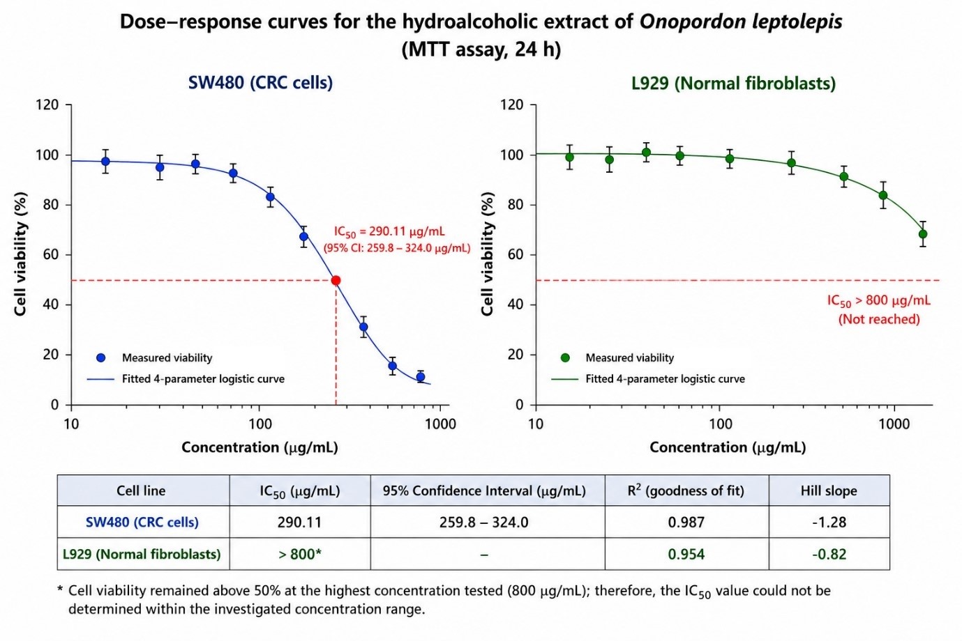
Concentration–response cytotoxicity profiles of
Comparative Effects of Onopordum leptolepis Extract on CRC Cell and Fibroblast Viability
Direct comparison of cell viability between SW480 CRC cells and L929 fibroblasts following 24 h of extract exposure across increasing concentrations (12.5–800 µg/mL) is presented in Figure 2. In SW480 cells, low extract concentrations (12.5–50 µg/mL) induced a modest but statistically significant decrease in viability (88–92% vs. 100% in control, p < 0.05). Substantial cytotoxicity occurred at intermediate concentrations of 100 and 200 µg/mL, where viability dropped significantly to approximately 70% and 55%, respectively (p < 0.001 vs. control). At 400 and 800 µg/mL, cell survival further declined to 48% and 32%, respectively (p < 0.001 vs. control), representing more than a two-thirds reduction in metabolic activity. Conversely, L929 fibroblasts maintained near-baseline viability at concentrations up to 200 µg/mL (p > 0.05 vs. control), with only a slight reduction at 400 µg/mL (approximately 88% viability, p < 0.01) and a moderate reduction at 800 µg/mL (around 75% viability, p < 0.001) (Figure 2).
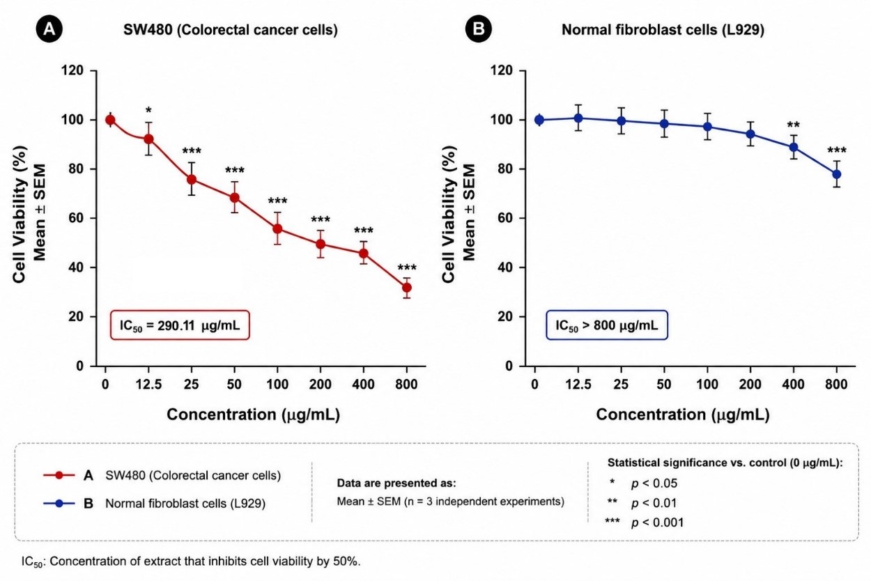
Comparative metabolic viability of SW480 colorectal cancer cells and L929 fibroblasts following treatment with
Induction of Apoptotic DNA Fragmentation in SW480 Cells
To determine whether extract-induced cytotoxicity was associated with programmed cell death, apoptotic DNA fragmentation was quantified using the DPA colorimetric assay (Figure 3). Treatment of SW480 cells with O. leptolepis extract for 24 h led to a robust, dose-dependent increase in DNA fragmentation. Baseline fragmentation in untreated control cells was minimal (<5%). Exposure to low concentrations (12.5 and 25 µg/mL) resulted in a modest elevation of fragmentation to 8–12% (p < 0.05). A marked and statistically significant increase was observed at 50 and 100 µg/mL, where fragmentation rose to approximately 18% and 30%, respectively (p < 0.001). At 200 µg/mL, apoptosis reached nearly 42% (p < 0.001), representing more than an eight-fold increase relative to controls. Higher concentrations (400 and 800 µg/mL) elicited the most pronounced apoptotic responses, with fragmentation levels reaching approximately 48% and nearly 60%, respectively (both p < 0.001) (Figure 3).
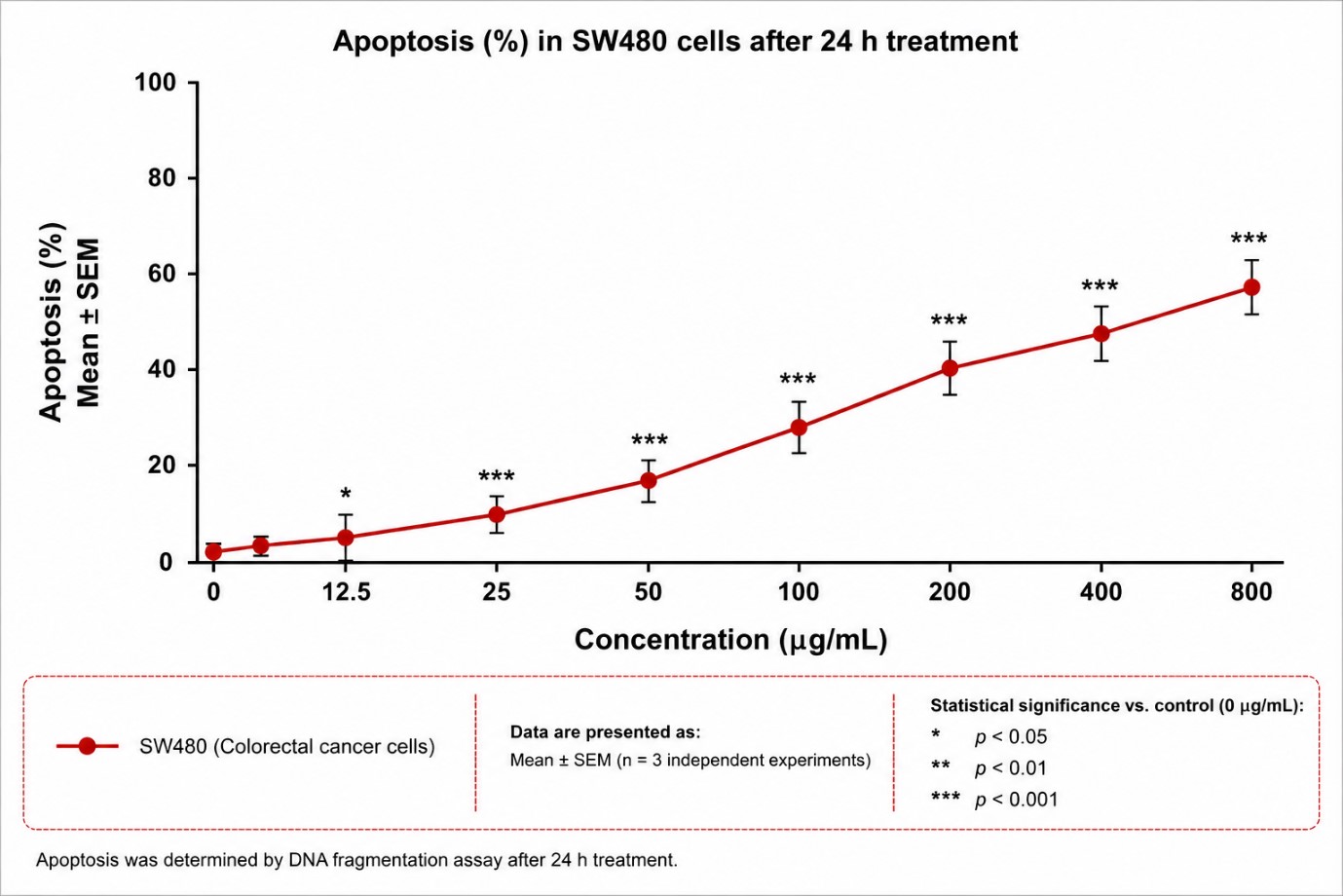
Concentration-dependent induction of apoptotic DNA fragmentation in SW480 cells by
Suppression of Notch Pathway Gene Expression in SW480 Cells
To examine the molecular mechanisms associated with the anticancer effects of O. leptolepis, relative mRNA expression levels of key Notch signaling components (Notch1, Jagged1, and c-Myc) were analyzed by qRT-PCR following 24 h of treatment (Figure 4). In untreated control cells, all three genes displayed stable baseline expression levels (normalized to 1.00 ± 0.10). Exposure to O. leptolepis extract resulted in significant transcriptional downregulation of Notch1 to 0.68 ± 0.12 (p < 0.001 vs. control) and Jagged1 to 0.63 ± 0.11 (p < 0.001 vs. control). Concurrently, expression of the downstream oncogenic effector c-Myc was significantly decreased to 0.52 ± 0.14 (p < 0.001 vs. control) (Figure 4).
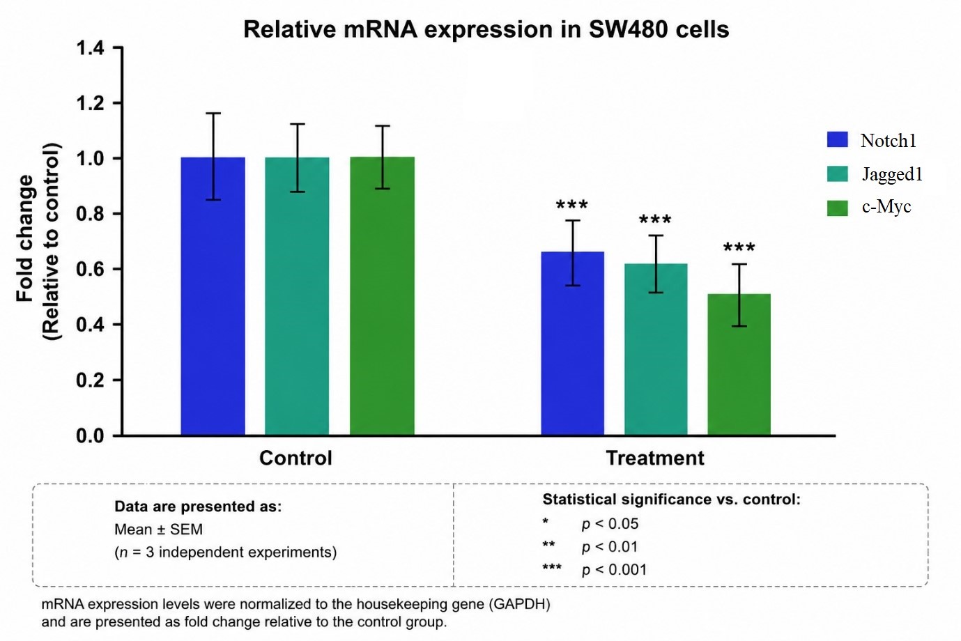
Transcriptional downregulation of Notch signaling components in SW480 cells treated with
Downregulation of Notch Pathway Protein Expression in SW480 Cells
Consistent with the observed transcriptional changes, quantitative ELISA demonstrated a significant reduction in cellular protein levels of Notch1, Jagged1, and c-Myc in SW480 cells following 24 h of extract treatment (Figure 5). Compared with untreated control cells (normalized to 1.0-fold), exposure to O. leptolepis extract significantly decreased Notch1 protein levels to approximately 0.80-fold, Jagged1 to 0.65-fold, and c-Myc to 0.60-fold (all p < 0.001 vs. control) (Figure 5).
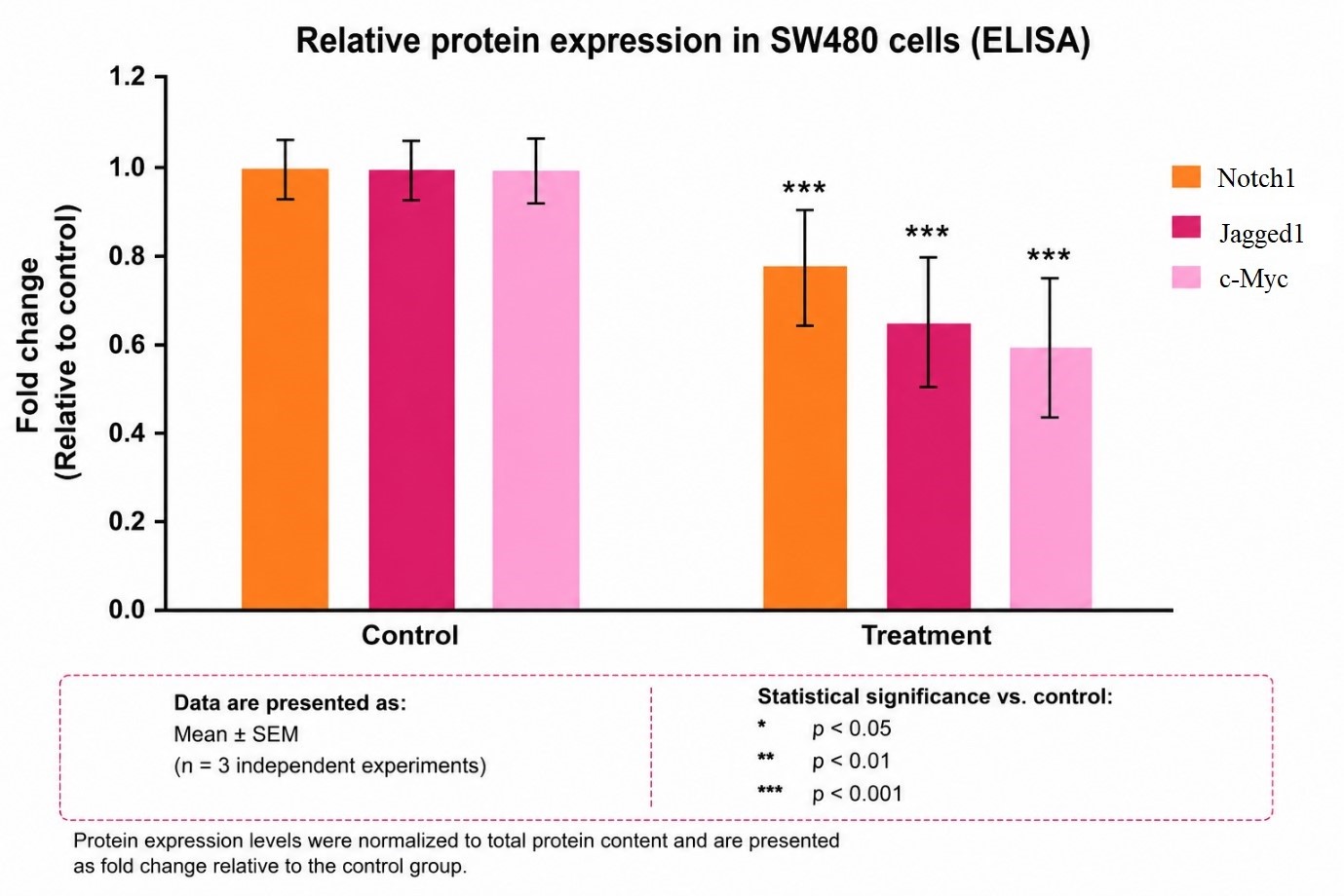
Suppression of Notch1, Jagged1, and c-Myc cellular protein levels in SW480 cells by
Inhibition of SW480 Cell Migration by Onopordum leptolepis Extract
The effect of O. leptolepis extract on cell motility was evaluated using the scratch wound-healing assay (Figure 6). Untreated control SW480 cells exhibited vigorous collective migration, achieving 45–50% scratch area closure after 24 h. In contrast, treatment with O. leptolepis extract significantly impeded cell migration, reducing wound closure to 20–25% (p < 0.001 vs. control), demonstrating potent antimigratory activity in vitro (Figure 6).
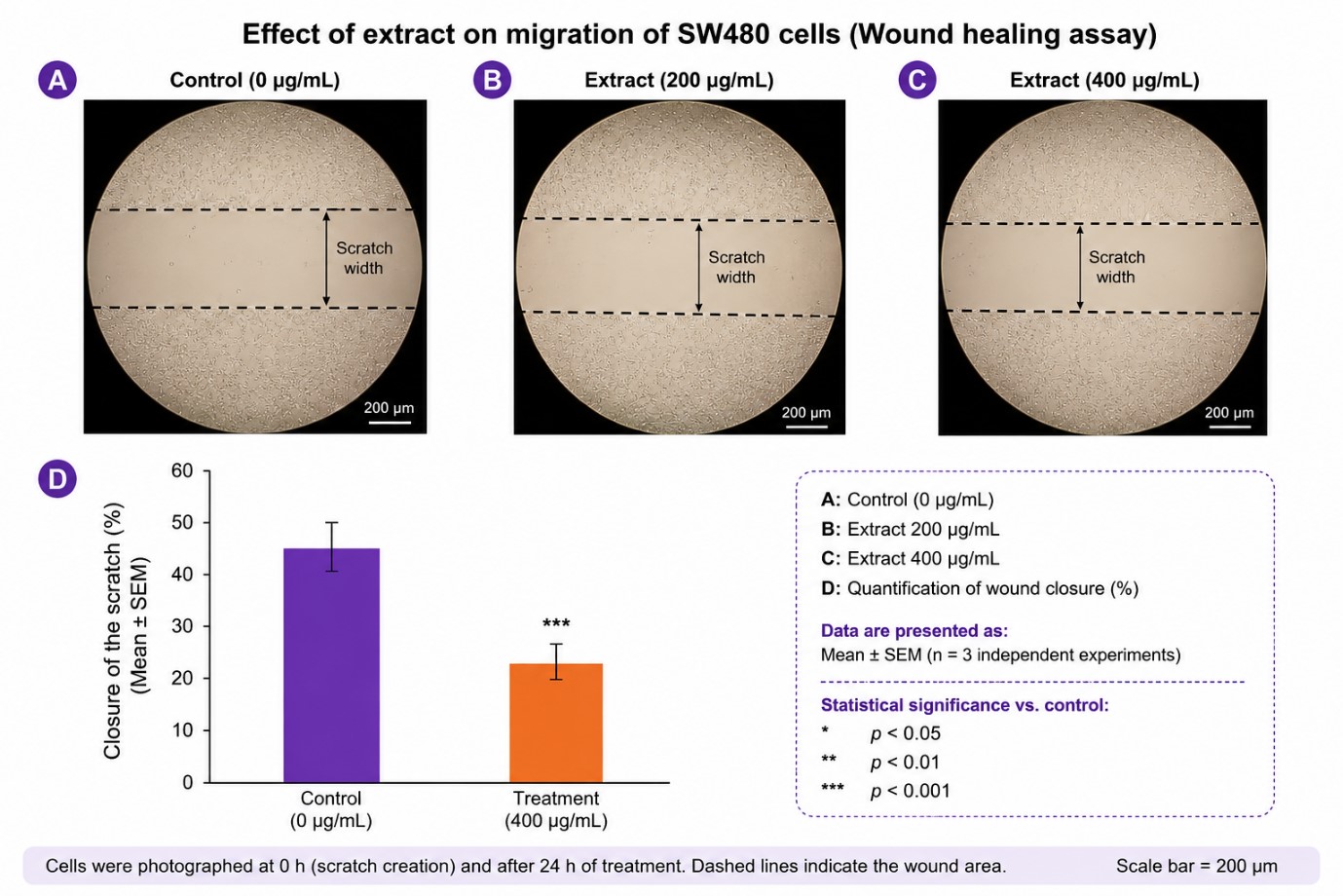
Attenuation of SW480 colorectal cancer cell migration by
Inhibition of SW480 Cell Invasion by Onopordum leptolepis Extract
The impact of O. leptolepis extract on cell invasiveness was assessed using Matrigel-coated Transwell invasion chambers (Figure 7). In untreated controls, robust cellular invasion through the reconstituted basement membrane was observed, corresponding to an optical density (OD) of 0.95–1.0. Exposure to O. leptolepis extract significantly decreased the absorbance of extracted crystal violet to approximately 0.80 (p < 0.001 vs. control), reflecting a substantial reduction in the number of invading cancer cells (Figure 7).
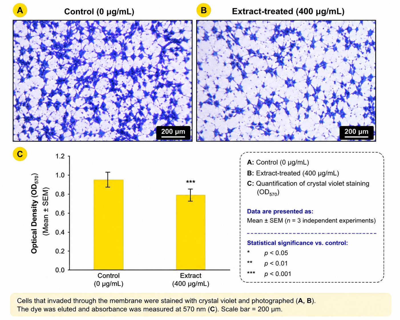
Inhibition of SW480 colorectal cancer cell invasion through Matrigel by
Discussion
To our knowledge, the present study provides the first evidence demonstrating that the 70% ethanol hydroalcoholic extract of Onopordum leptolepis exhibits potent in vitro antiproliferative, pro-apoptotic, antimigratory, and anti-invasive activities against SW480 colorectal cancer cells, accompanied by significant downregulation of Notch1, Jagged1, and c-Myc expression at both the transcript and protein levels. Although the modulation of Notch signaling has been established for several other botanical compounds, the antineoplastic potential of O. leptolepis in colorectal cancer has not been previously described24.
Our findings revealed that O. leptolepis extract induced a concentration-dependent reduction in SW480 cell viability (IC = 290.11 µg/mL) while exhibiting markedly lower cytotoxicity toward non-tumorigenic L929 fibroblasts (IC > 800 µg/mL). This differential response indicates a favorable index of selectivity toward malignant cells under the tested in vitro conditions. Growth inhibition was accompanied by a robust, dose-dependent induction of apoptosis, as evidenced by significant DNA fragmentation reaching nearly 60% at 800 µg/mL. These results suggest that the cytotoxic action of the extract is mediated, at least in part, by the activation of programmed cell death pathways.
The concentrations evaluated in this study (12.5–800 µg/mL) were selected to define the full concentration–response relationship of the crude extract during initial in vitro screening. While high in vitro concentrations (up to 800 µg/mL) are standard in preliminary botanical evaluations, such systemic levels may not be directly achievable in vivo due to complex pharmacokinetic parameters, including gastrointestinal absorption, first-pass hepatic metabolism, tissue distribution, and rapid clearance. Consequently, these findings should be interpreted as proof-of-concept evidence of biological activity in vitro rather than direct predictors of clinical efficacy. Future investigations focusing on bioassay-guided fractionation, active constituent isolation, pharmacokinetic profiling, and in vivo xenograft models are required to establish translational relevance and therapeutically achievable doses.
Aberrant activation of the Notch signaling pathway plays a pivotal role in the initiation, progression, and metastasis of various malignancies, including colorectal carcinoma25, prostate carcinoma26, and breast carcinoma27. In CRC, elevated expression of Notch1, its ligand Jagged1, and downstream target genes such as c-Myc, Hes-1, DLL4, and NF-κB contributes to sustained proliferative signaling, evasion of apoptosis, maintenance of cancer stem cell populations, and epithelial–mesenchymal transition (EMT)28. Several well-characterized phytochemicals, such as curcumin, resveratrol, quercetin, epigallocatechin-3-gallate (EGCG), berberine, and thymoquinone, have been reported to exert anticancer effects by inhibiting Notch signaling components. Consistent with these observations, our study demonstrated that O. leptolepis extract significantly decreased the mRNA and protein levels of Notch1, Jagged1, and c-Myc in SW480 cells, suggesting that downregulation of this signaling axis may represent an important mechanistic contributor to its antitumor properties.
In addition to suppressing proliferation and inducing apoptosis, O. leptolepis extract significantly reduced SW480 cell motility in wound-healing assays and restricted invasion through Matrigel barriers. Because Notch signaling is known to drive tumor cell motility and invasiveness by orchestrating cytoskeletal remodeling, matrix metalloproteinase expression, and EMT progression, the observed antimigratory and anti-invasive actions of the extract may stem, in part, from Notch pathway suppression.
An interesting aspect of our molecular findings is that the reduction in protein levels of Notch1, Jagged1, and c-Myc (0.60–0.80-fold of control) was somewhat less pronounced than their corresponding transcriptional downregulation (0.52–0.68-fold of control). Such differences between transcript abundance and protein levels are common in biological systems and can be attributed to multiple post-transcriptional and translational regulatory mechanisms, including mRNA stability, translation efficiency, post-translational modifications, and protein turnover rates. Furthermore, the 24-h treatment window employed here may have been sufficient to substantially inhibit gene transcription while only partially depleting pre-existing cellular protein pools, particularly for proteins with longer intracellular half-lives. Time-course studies evaluating multiple exposure durations and protein turnover kinetics will be valuable to delineate the temporal dynamics of pathway inhibition.
Our findings align with and expand upon previous phytochemical and pharmacological investigations of Onopordum leptolepis. Mirzaei et al. investigated the aerial parts of O. leptolepis using GC-MS analysis, identifying 27 chemical constituents, including alkaloids, cyanogenic glycosides, tannins, and steroids, and demonstrated antiproliferative effects against CACO2 colorectal cancer cells via MTT and NBT assays29,30. Similarly, Valizadeh et al. evaluated various solvent extracts of vegetative and floral parts of O. leptolepis, reporting that hydroalcoholic extracts possessed the highest free radical scavenging and ferric reducing antioxidant power (FRAP) activities9,30. Furthermore, Njeh et al. isolated active flavonoids (including hispidulin and dehydromelitensin derivatives) from related Onopordum species (O. espinae) and demonstrated significant antimicrobial and biological activities31. Collectively, these studies suggest that the rich phytoconstituent profile of O. leptolepis, particularly its flavonoid, alkaloid, and phenolic content, underpins its broad antioxidant and antineoplastic properties.
Although our results demonstrate a strong association between O. leptolepis treatment and downregulation of the Notch1/Jagged1/c-Myc axis, the present study does not establish a direct causal molecular target. At higher concentrations, crude botanical extracts may also exert non-specific cellular stress, membrane perturbation, or mitochondrial effects that contribute to cytotoxicity. Nonetheless, the marked selectivity observed between SW480 cells and normal L929 fibroblasts, coupled with coordinated gene and protein downregulation, supports target-specific biological activity beyond generalized toxicity.
Limitations
This study has several limitations that should be acknowledged. First, the antitumor evaluation was conducted exclusively in a single colorectal cancer cell line (SW480); validation across additional CRC lines representing distinct genetic backgrounds (e.g., HCT116, HT-29, DLD-1) and primary patient-derived cells is necessary. Second, the observed link between extract treatment and Notch pathway downregulation remains associative; genetic gain-of-function (overexpression) or rescue experiments will be required to confirm direct causality. Third, the study utilized a crude 70% ethanol extract without bioassay-guided fractionation or chromatographic isolation of individual active constituents. Consequently, whether the observed activities are driven by single compounds or synergistic phytochemical interactions remains to be elucidated. Finally, as an in vitro investigation, these findings cannot directly predict in vivo bioavailability, pharmacokinetics, systemic toxicity, or therapeutic efficacy, necessitating future preclinical animal model evaluations.
Conclusion
In conclusion, the 70% ethanol hydroalcoholic extract of Onopordum leptolepis exhibits selective in vitro antiproliferative, pro-apoptotic, antimigratory, and anti-invasive activities against SW480 colorectal cancer cells. These phenotypic antitumor effects are accompanied by significant downregulation of the Notch1/Jagged1/c-Myc signaling axis at both the mRNA and protein levels. While these findings highlight O. leptolepis as a promising botanical candidate for colorectal cancer therapeutics, further research encompassing phytochemical standardization, isolation of active constituents, causal pathway validation, and in vivo evaluations is warranted to fully establish its translational potential.
Abbreviations
ANOVA: Analysis of variance; cDNA: Complementary DNA; CRC: Colorectal carcinoma; DMEM: Dulbecco’s Modified Eagle Medium; DMSO: Dimethyl sulfoxide; DPA: Diphenylamine; EDTA: Ethylenediaminetetraacetic acid; EGCG: Epigallocatechin-3-gallate; ELISA: Enzyme-linked immunosorbent assay; EMT: Epithelial–mesenchymal transition; FBS: Fetal bovine serum; FRAP: Ferric reducing antioxidant power; GAPDH: Glyceraldehyde-3-phosphate dehydrogenase; GC-MS: Gas chromatography–mass spectrometry; IARC: International Agency for Research on Cancer; IC: Half-maximal inhibitory concentration; MTT: 3-(4,5-Dimethylthiazol-2-yl)-2,5-diphenyltetrazolium bromide; NBT: Nitroblue tetrazolium; NICD: Notch intracellular domain; OD: Optical density; PBS: Phosphate-buffered saline; qRT-PCR: Quantitative reverse transcription polymerase chain reaction; RPMI: Roswell Park Memorial Institute; SEM: Standard error of the mean; TCA: Trichloroacetic acid; TMB: 3,3′,5,5′-Tetramethylbenzidine.
Acknowledgments
None.
Author’s Contributions
Conceptualization: F.K-H.; Methodology: M.P.; Software: I.R.; Data curation: N.S.; Investigation: C.J.; Validation: M.P.; Formal analysis: F.K-H.; Supervision: F.K-H.; Funding acquisition: F.K-H.; Visualization: M.P.; Project administration: F.K-H.; Resources: F.K-H.; Writing (original draft): A.A.; Writing (review & editing): A.A. All authors read and approved the final manuscript.
Funding
This study was supported by a research grant from the Research Council of Kermanshah University of Medical Sciences (Grant No. 4030466).
Availability of Data and Materials
The datasets used and analyzed during the current study are available from the corresponding author upon reasonable request.
Ethics Approval and Consent to Participate
This study was approved by the Ethics Committee of Kermanshah University of Medical Sciences (Ethical approval code: IR.KUMS.MED.REC.1402.388). For this in vitro study using established cell lines, consent to participate was not applicable.
Consent for Publication
Not applicable.
Declaration of Generative AI and AI-Assisted Technologies in the Writing Process
None.
Competing Interests
The authors declare that they have no competing interests.