


Impact of Super Growth Differentiation Factor 9 Supplementation in a Biphasic In Vitro Maturation Culture System on Embryo Morphokinetic Development: A Secondary Analysis of a Sibling-Oocyte Cohort

- Faculty of Biology and Biotechnology, University of Science, Ho Chi Minh City, Vietnam
- Vietnam National University, Ho Chi Minh City, Vietnam
- IVF Quoc Anh, Quoc Anh General Hospital, Ho Chi Minh City, Vietnam
- HOPE Research Center, My Duc Hospital, Ho Chi Minh City, Vietnam
- IVFMD, My Duc Hospital, Ho Chi Minh City, Vietnam
- IVFMD Phu Nhuan, My Duc Phu Nhuan Hospital, Ho Chi Minh City, Vietnam
- Department of Obstetrics and Gynecology, University of Medicine and Pharmacy at Ho Chi Minh City, Ho Chi Minh City, Vietnam
Abstract
Introduction: In vitro maturation (IVM) is a safer, cost-effective alternative to conventional in vitro fertilization (IVF), particularly for individuals with polyendocrine metabolic ovarian syndrome (PMOS). However, clinical adoption of IVM remains constrained by reduced oocyte developmental competence and suboptimal reproductive outcomes, in part due to asynchrony between nuclear and cytoplasmic maturation. Capacitation in vitro maturation (CAPA-IVM) was developed to improve oocyte competence by introducing a pre-maturation phase; nonetheless, clinical outcomes remain inferior to conventional IVF, underscoring the need for further culture optimization. Oocyte-secreted factors (OSFs), specifically growth differentiation factor 9 (GDF9) and bone morphogenetic protein 15 (BMP15), are crucial paracrine regulators of cumulus–oocyte communication but present translational challenges in clinical practice. Super-GDF9 (sGDF9), a bioengineered GDF9 variant with markedly enhanced bioactivity, represents a promising candidate. This study evaluated human embryo morphokinetic development following CAPA-IVM with or without sGDF9 supplementation using continuous time-lapse imaging.
Methods: This was a secondary analysis of a prospective sibling-oocyte clinical trial (ClinicalTrials.gov identifier: NCT06766604) in women aged 18–35 years with PMOS undergoing CAPA-IVM. Sibling cumulus–oocyte complexes (COCs) were allocated to standard CAPA-IVM culture media (control group) or media supplemented with 50 ng/mL sGDF9 during both the pre-IVM and maturation phases. In total, 135 annotated embryos (85 control and 50 sGDF9) from nine patients were monitored using a continuous time-lapse imaging incubator. Morphokinetic parameters, including cleavage timings (t2PN to t8), cell cycle intervals, developmental milestones (compaction and blastulation), and abnormal cleavage patterns, were compared between groups. Linear and generalized linear mixed-effects models with a patient-level random intercept were applied to account for embryo clustering within patients.
Results: Early cleavage morphokinetics (t2PN through eight-cell stage [t8]) were comparable between the control and sGDF9 groups. In unadjusted comparisons, the time to full blastocyst expansion (tB) was significantly prolonged in the sGDF9 group compared with controls (120.0 ± 11.6 vs. 100.0 ± 7.2 h; p = 0.036; analyzed in a subset of n = 16 blastocysts). Consistently, multivariable mixed-effects models demonstrated no significant differences across early morphokinetic parameters (all p > 0.05), whereas tB was significantly delayed following sGDF9 supplementation (adjusted mean difference: 18.98 h; 95% CI: 7.88–30.08; nominal p = 0.002; Benjamini–Hochberg adjusted p = 0.030). A non-significant trend toward longer time to the 8-cell stage was also observed in the sGDF9 group (adjusted mean difference: 5.25 h; 95% CI: −0.45 to 10.94; p = 0.070). Rates of abnormal cleavage patterns did not differ significantly between groups (p = 0.117).
Conclusion: In this secondary analysis of a sibling-oocyte cohort, sGDF9 supplementation during CAPA-IVM was not associated with significant alterations in early embryo cleavage morphokinetics or abnormal cleavage patterns. Although blastocyst expansion was delayed in a small subset of embryos reaching the blastocyst stage, overall developmental kinetics were broadly comparable between groups. These findings suggest that the benefits of sGDF9 observed in conventional IVM may not directly translate to biphasic CAPA-IVM systems.
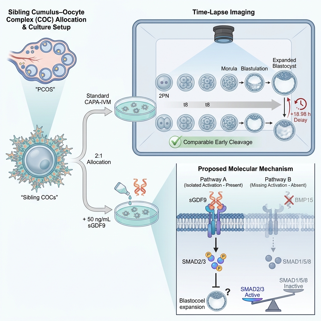
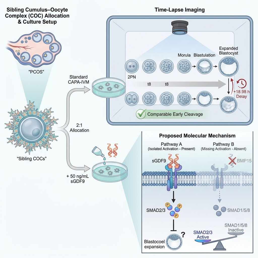
Graphical abstract
Introduction
In vitro maturation (IVM) is an assisted reproductive technology in which immature oocytes retrieved from small antral follicles undergo maturation in vitro prior to fertilization.1,2 Compared with conventional in vitro fertilization (IVF), IVM offers notable clinical advantages, including minimal or no gonadotropin stimulation, lower overall treatment costs, and complete elimination of the risk of ovarian hyperstimulation syndrome (OHSS).3,4,5,6 These characteristics render IVM particularly advantageous for women with polycystic ovary syndrome (PCOS)—recently recognized under international consensus as polyendocrine metabolic ovarian syndrome (PMOS)7—as well as individuals with high ovarian reserve and patients requiring urgent fertility preservation. Nevertheless, the widespread clinical adoption of IVM remains constrained by lower blastocyst yields and inferior cumulative live birth rates compared with conventional IVF.8,9
A principal factor compromising IVM oocyte developmental competence is the desynchronization between nuclear and cytoplasmic maturation during in vitro culture. In vivo, oocyte meiotic arrest is sustained within the ovarian follicle while the ooplasm progressively accumulates maternal transcripts, proteins, and metabolic organelles essential for fertilization and subsequent embryogenesis. Rapid spontaneous meiotic resumption upon retrieval disrupts this balance. To address this limitation, a biphasic culture strategy termed capacitation-IVM (CAPA-IVM) was developed.10 By incorporating a pre-IVM phase that maintains elevated cyclic adenosine monophosphate (cAMP) levels and sustains gap-junction communication between the oocyte and surrounding cumulus cells, CAPA-IVM promotes coordinated cytoplasmic and nuclear maturation.11 Although CAPA-IVM significantly improves oocyte developmental competence compared with standard IVM, usable blastocyst numbers and cumulative pregnancy rates remain lower than those achieved with conventional IVF, underscoring the necessity for further optimization of the in vitro maturation milieu.8,9
Oocyte developmental competence is governed by dynamic bidirectional paracrine signaling between the oocyte and cumulus cells, mediated predominantly by oocyte-secreted factors (OSFs). Among these, growth differentiation factor 9 (GDF9) and bone morphogenetic protein 15 (BMP15) are pivotal regulators of follicular development and granulosa cell function, acting through downstream SMAD signaling cascades.12,13,14 Exogenous supplementation of IVM media with OSFs has been shown to improve oocyte maturation, fertilization, and embryonic developmental capacity in both human and animal models.15,16,17 Notably, GDF9 and BMP15 form a heterodimer, termed cumulin, that exhibits dramatically higher bioactivity in activating granulosa cell signaling than either homodimer alone.8,14 However, the translational utility of recombinant cumulin is hindered by substantial biochemical challenges related to production yield and purification stability. To circumvent these constraints, a bioengineered variant of human GDF9, designated Super-GDF9 (sGDF9), was developed. This engineered factor exhibits over 1,000-fold greater potency than wild-type GDF9 in activating SMAD2/3 signaling in granulosa cells.14,18 Although sGDF9 operates primarily as a homodimeric GDF9 mimic and activates the SMAD2/3 pathway without concomitant BMP15-mediated SMAD1/5/8 activation, its enhanced bioactivity was hypothesized to strengthen oocyte–cumulus communication and establish a more physiologically supportive follicular environment during IVM.14,18 While supplementation with cumulin or sGDF9 enhances oocyte competence and blastocyst formation in conventional IVM systems,8 its specific impact within biphasic systems such as CAPA-IVM remains poorly defined.
Recent advances in time-lapse imaging technology enable continuous, non-invasive observation of preimplantation embryo development under undisturbed, highly controlled culture conditions. This platform permits precise characterization of morphokinetic parameters—such as the exact timing of early cleavage divisions, cell cycle durations, synchronization intervals, and abnormal division behaviors—that are closely correlated with developmental competence, chromosomal constitution, and implantation potential.19 Consequently, morphokinetic assessment serves as a sensitive tool to evaluate how specific culture media modifications during oocyte maturation influence downstream embryo development.
Given the critical role of OSFs in orchestrating cumulus–oocyte communication and the enhanced potency of sGDF9, rigorous evaluation is needed to determine whether supplementing CAPA-IVM culture with sGDF9 confers developmental benefits during subsequent embryogenesis. Therefore, this study aimed to investigate the impact of sGDF9 supplementation during the pre-IVM and IVM phases of the CAPA-IVM system on subsequent human embryo morphokinetics and cleavage dynamics using time-lapse imaging technology.
Materials and Methods
This study was a secondary analysis of a prospective, sibling-oocyte clinical trial evaluating CAPA-IVM in women with PMOS, previously described by Vuong et al. (2026).20 The original study protocol was reviewed and approved by the Institutional Ethics Committee of My Duc Hospital (approval reference: 12/24/DD-BVMD; November 25, 2024) and prospectively registered at ClinicalTrials.gov (identifier: NCT06766604). The study was conducted in accordance with the Declaration of Helsinki and the International Council for Harmonisation–Good Clinical Practice (ICH-GCP) guidelines. All participants received comprehensive counseling and provided written informed consent prior to enrollment.
Study Participants and Design
Eligible participants were women aged <35 years diagnosed with PMOS according to the Rotterdam criteria established by the European Society of Human Reproduction and Embryology (ESHRE) and the American Society for Reproductive Medicine (ASRM) who were scheduled for CAPA-IVM treatment. Inclusion criteria required the retrieval of ≥30 cumulus–oocyte complexes (COCs) in a single retrieval cycle and written consent to allocate ≥10 COCs to the sGDF9 experimental group for research purposes. Exclusion criteria comprised uterine anomalies, uncorrected ovarian pathology, and severe male factor infertility (defined as surgically retrieved sperm or severe oligoasthenoteratozoospermia).
Following oocyte retrieval, retrieved sibling COCs were randomly allocated into two study groups in a 2:1 ratio: the control group (n = 200 COCs) and the sGDF9 supplementation group (n = 100 COCs) (Figure 1). All laboratory protocols, culture media baselines, and environmental conditions were strictly standardized between groups. Throughout the pre-IVM and IVM stages, COCs were cultured under mineral oil overlay at 37°C in a humidified atmosphere of 7% CO and 20% O using a Planer BT37 incubator (Origio, Måløv, Denmark).3 Following in vitro maturation, denudation, and fertilization via intracytoplasmic sperm injection (ICSI), resulting zygotes were cultured in a multi-well time-lapse imaging incubator (CCM-iBIS, Astec, Fukuoka, Japan) under reduced oxygen conditions (5% O, 5% CO, and 90% N) at 37°C under oil to monitor embryonic progression.
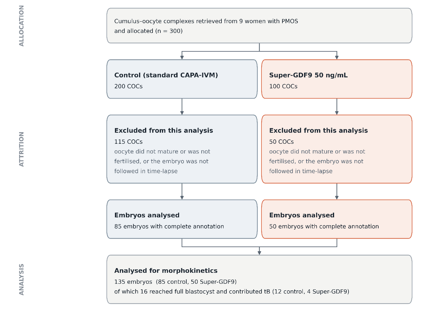
Flow diagram of cumulus–oocyte complex allocation, developmental attrition, and embryo morphokinetic analysis. A total of 300 cumulus–oocyte complexes (COCs) were retrieved from nine women aged <35 years diagnosed with polyendocrine metabolic ovarian syndrome (PMOS) undergoing capacitation in vitro maturation (CAPA-IVM). Retrieved sibling oocytes were allocated in a 2:1 ratio to either standard CAPA-IVM culture media (control group, n = 200) or media supplemented with 50 ng/mL super growth differentiation factor 9 (sGDF9 group, n = 100) during both the pre-IVM and IVM phases. Embryos developing following insemination were cultured under continuous time-lapse monitoring (CCM-iBIS). Following exclusion of oocytes that failed to mature, unfertilized oocytes, or embryos without complete time-lapse annotations (115 in the control group and 50 in the sGDF9 group), 135 embryos (85 control and 50 sGDF9) were included in the primary morphokinetic evaluation. A subset of 16 embryos (12 control and 4 sGDF9) successfully reached the fully expanded blastocyst stage and contributed to time-to-blastocyst (tB) analyses. CAPA-IVM, capacitation in vitro maturation; COCs, cumulus–oocyte complexes; PMOS, polyendocrine metabolic ovarian syndrome; sGDF9, super growth differentiation factor 9; tB, time to full blastocyst development.
In this secondary analysis, 135 embryos derived from 300 COCs across nine patients with complete morphokinetic annotations were evaluated (85 embryos in the control group and 50 embryos in the sGDF9 group). Embryos in the sGDF9 group were exposed to 50 ng/mL sGDF9 throughout both the pre-IVM and maturation phases, whereas control embryos were cultured in standard unsupplemented CAPA-IVM media. The 50 ng/mL dosage was selected based on established preclinical murine titration data and the biological hypothesis that endogenous GDF9 and BMP15 translational activities decline from the germinal vesicle to the metaphase II stage.8,21 Unlike native recombinant GDF9, which is typically administered at concentrations of 100–200 ng/mL, sGDF9 exhibits >1,000-fold greater SMAD2/3 signaling bioactivity.8,17,22 Consequently, 50 ng/mL was considered optimal to achieve robust biological receptor activation without excessive cellular overstimulation, following the dosing rationale described by Stocker et al. (2020).14
Morphokinetic Evaluation
Embryo morphokinetics were assessed using the time-lapse monitoring system, enabling continuous, non-invasive digital imaging without disturbing incubation conditions. Automated image acquisition was performed at 10-minute intervals across multiple focal planes. All morphokinetic annotations were performed by a single senior embryologist with more than five years of experience in time-lapse embryo assessment, using standardized criteria. The assessment was not blinded to study allocation.
Kinetic milestones were defined as hours post-insemination (hours post-ICSI), including: time to pronuclear appearance (t2PN), time to pronuclear fading (tPNf), and timings of cleavage divisions from the 2-cell to 8-cell stages (t2, t3, t4, t5, t6, t7, and t8). Cell cycle dynamics were assessed by calculating the duration of the second cell cycle (cc2 = t3 − t2), the synchronicity of the second division (s2 = t4 − t3), and the synchronicity of the third division (s3 = t8 − t5), according to consensus guidelines established by Basile et al. (2014)23 and the ESHRE Working Group on Time-Lapse Technology.19
In addition, abnormal cleavage patterns were systematically recorded: (1) direct cleavage (dc), defined as an abnormally rapid division yielding three blastomeres within <5 hours, subcategorized into dc1 (direct cleavage during the first cell cycle, encompassing direct transition from 1 to 3 blastomeres or an interval between t2 and t3 of <5 hours) and dc2 (direct cleavage during subsequent cycles); (2) reverse cleavage (rc), defined as blastomere fusion or failure of cytokinesis resulting in a reduced cell count following division; and (3) multinucleated blastomeres at the 2- or 4-cell stage.
For extended culture, late preimplantation milestones were annotated: time to initiation of compaction (tSC), time to full compaction/morula stage (tC/tM), time to initiation of blastulation/cavitation (tSB), and time to full blastocyst expansion (tB).
Statistical Analysis
Continuous variables were assessed for distributional normality using the Shapiro–Wilk test and visual inspection of Q–Q plots. Normally distributed variables are expressed as mean ± standard deviation (SD), and non-normally distributed data are reported as median (interquartile range [IQR]). Unadjusted comparisons between groups were performed using the two-tailed Student’s t-test for normally distributed variables and the Wilcoxon rank-sum test for skewed data. Categorical variables are presented as absolute frequencies and percentages and were evaluated using Fisher’s exact test.
To rigorously account for the hierarchical clustering of multiple embryos originating from the same patient (non-independence of sibling-oocyte observations), multivariable mixed-effects models specifying a random intercept for patient were implemented. Continuous morphokinetic outcomes were modeled using linear mixed-effects models (lme4::lmer formulation: outcome ~ group + (1 | patient)), fitted via Restricted Maximum Likelihood (REML). Satterthwaite approximations were employed to estimate denominator degrees of freedom (lmerTest package), with results reported as adjusted mean differences, 95% confidence intervals (CI), and p-values. Binary outcomes, including abnormal cleavage patterns, were analyzed using generalized linear mixed-effects models (glmer) with the Laplace approximation and are reported as conditional odds ratios (OR) with Wald 95% CIs. Multiple testing across morphokinetic parameters was controlled using the Benjamini–Hochberg (BH) false discovery rate method. All analyses were conducted on available case data without imputation for missing milestones resulting from developmental arrest or Day 3 intervention. A two-sided p-value <0.05 was considered statistically significant. Statistical analyses were performed using R software version 4.5.1 (R Foundation for Statistical Computing, Vienna, Austria).
Results
Study Population and Cohort Attrition
A total of nine women diagnosed with PMOS were enrolled in the study, contributing 300 retrieved sibling COCs (200 allocated to standard CAPA-IVM control media and 100 allocated to sGDF9-supplemented media; Figure 1), as previously described by Vuong et al. (2026).20 Following oocyte maturation and fertilization, 165 COCs did not generate annotated embryos due to maturation failure, non-fertilization, or lack of continuous time-lapse annotation (115 in the control group and 50 in the sGDF9 group). Consequently, 135 annotated embryos (85 control and 50 sGDF9) from all nine patients were analyzed at the embryo level for preimplantation morphokinetics.
Embryo Morphokinetics
In unadjusted descriptive analyses, early cleavage timings (t2PN through t8), cell cycle durations (cc2), and division synchronicity (s2 and s3) demonstrated substantial overlap between the control and sGDF9 groups (Table 1; Figures 2 and 3), reflecting broadly equivalent early mitotic kinetics. Mean timings post-insemination were comparable between control and sGDF9 groups for t2PN (11.6 ± 2.8 vs. 12.3 ± 3.8 h; p = 0.292), t2 (28.4 ± 7.1 vs. 30.8 ± 11.0 h; p = 0.184), t3 (34.6 ± 11.8 vs. 34.0 ± 7.4 h; p = 0.715), t4 (37.8 ± 11.2 vs. 36.4 ± 8.2 h; p = 0.444), t5 (42.8 ± 11.3 vs. 42.3 ± 10.7 h; p = 0.812), and t8 (57.9 ± 12.8 vs. 62.2 ± 14.7 h; p = 0.178).
Unadjusted morphokinetic parameters and cleavage patterns of embryos derived from capacitation in vitro maturation with or without super-GDF9 supplementation.
| Parameter | Subgroup / Category | Control Group | Super-GDF9 Group | p-value | ||||
|---|---|---|---|---|---|---|---|---|
| Embryos (n) | Patients (n) | Value (Mean ± SD or n [%]) | Embryos (n) | Patients (n) | Value (Mean ± SD or n [%]) | |||
| t2PN (h) | All annotated embryos | 83 | 9 | 11.6 ± 2.8 | 44 | 9 | 12.3 ± 3.8 | 0.292 |
| t2 (h) | All annotated embryos | 81 | 9 | 28.4 ± 7.1 | 48 | 9 | 30.8 ± 11.0 | 0.184 |
| t3 (h) | All annotated embryos | 78 | 9 | 34.6 ± 11.8 | 47 | 9 | 34.0 ± 7.4 | 0.715 |
| t4 (h) | All annotated embryos | 76 | 9 | 37.8 ± 11.2 | 46 | 9 | 36.4 ± 8.2 | 0.444 |
| t5 (h) | All annotated embryos | 73 | 9 | 42.8 ± 11.3 | 45 | 9 | 42.3 ± 10.7 | 0.812 |
| t8 (h) | All annotated embryos | 58 | 9 | 57.9 ± 12.8 | 29 | 9 | 62.2 ± 14.7 | 0.178 |
| tSC (h) | All annotated embryos | 46 | 8 | 68.8 ± 11.4 | 18 | 7 | 76.2 ± 18.1 | 0.121 |
| tC (h) | All annotated embryos | 35 | 6 | 86.1 ± 14.0 | 9 | 4 | 92.5 ± 13.7 | 0.232 |
| tSB (h) | Reached full blastocyst (tSB & tB annotated) | 29 | 6 | 93.0 ± 8.9 (n = 12) | 9 | 6 | 107.0 ± 10.3 (n = 4) | — |
| Initiated but did not complete blastulation | 29 | 6 | 108.6 ± 8.5 (n = 17) | 9 | 6 | 105.5 ± 7.1 (n = 5) | — | |
| All embryos initiating blastulation | 29 | 6 | 102.1 ± 11.6 (n = 29) | 9 | 6 | 106.2 ± 8.1 (n = 9) | 0.256 | |
| tB (h) | Reached full blastocyst (tSB & tB annotated) | 12 | 6 | 100.5 ± 7.2 (n = 12) | 4 | 3 | 119.9 ± 11.6 (n = 4) | — |
| Initiated but did not complete blastulation | 12 | 6 | Not reached | 4 | 3 | Not reached | — | |
| All embryos forming full blastocyst | 12 | 6 | 100.5 ± 7.2 (n = 12) | 4 | 3 | 120.0 ± 11.6 (n = 4) | 0.036 | |
| cc2 (t3 − t2) (h) | Second cell cycle duration | 78 | 9 | 6.6 ± 8.5 | 47 | 9 | 4.5 ± 4.5 | 0.074 |
| s2 (t4 − t3) (h) | Second cleavage synchronicity | 76 | 9 | 3.7 ± 4.5 | 45 | 9 | 2.7 ± 4.3 | 0.201 |
| s3 (t8 − t5) (h) | Third cleavage synchronicity | 58 | 9 | 16.3 ± 12.3 | 29 | 9 | 19.7 ± 11.5 | 0.203 |
| No abnormal cleavage | Normal cleavage pattern | 85 | 9 | 36/85 (42.4%) | 50 | 9 | 14/50 (28.0%) | 0.117 |
| Direct cleavage 1 (dc1) | First division abnormality (≤5 h) | 85 | 9 | 39/85 (45.9%) | 50 | 9 | 31/50 (62.0%) | 0.077 |
| Direct cleavage 2 (dc2) | Subsequent division abnormality | 85 | 9 | 10/85 (11.8%) | 50 | 9 | 4/50 (8.0%) | 0.571 |
| Reverse cleavage (rc) | Blastomere fusion / reduction | 85 | 9 | 0/85 (0.0%) | 50 | 9 | 1/50 (2.0%) | 0.370 |
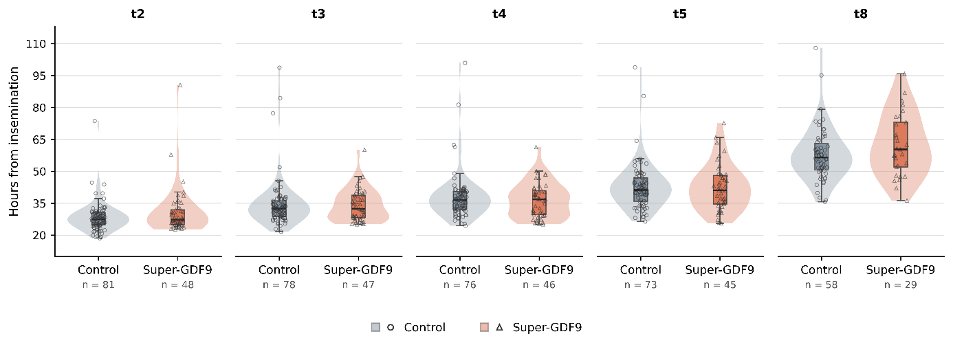
Embryo cleavage timings across early cleavage stages (t2 to t8) following capacitation in vitro maturation with or without super-GDF9 supplementation. Preimplantation morphokinetic timings from the 2-cell stage (t2) through the 8-cell stage (t8) were tracked continuously using time-lapse imaging (CCM-iBIS) following insemination of sibling oocytes matured in standard CAPA-IVM media (Control) or media supplemented with 50 ng/mL super-GDF9 (Super-GDF9). Timings are expressed as hours post-insemination. Early cleavage timings showed substantial overlap and were statistically comparable between the control and super-GDF9 groups across all stages (t2, t3, t4, t5, and t8; all unadjusted and adjusted p > 0.05). A non-significant trend toward prolonged cleavage timing was observed at the 8-cell stage in the super-GDF9 group (adjusted mean difference, 5.25 h; p = 0.070). Violin plots depict the distribution density of cleavage timings; embedded box plots represent the median (horizontal black line) and interquartile range (IQR; 25th to 75th percentiles); whiskers indicate 1.5 × IQR. Individual data points represent single embryos (gray circles, control; orange triangles, super-GDF9). Sample sizes (n) for each stage represent the number of embryos reaching that milestone and are indicated beneath each plot. sGDF9, super growth differentiation factor 9; t2, t3, t4, t5, t8, time from insemination to cleavage to 2-, 3-, 4-, 5-, and 8-cell stages, respectively.
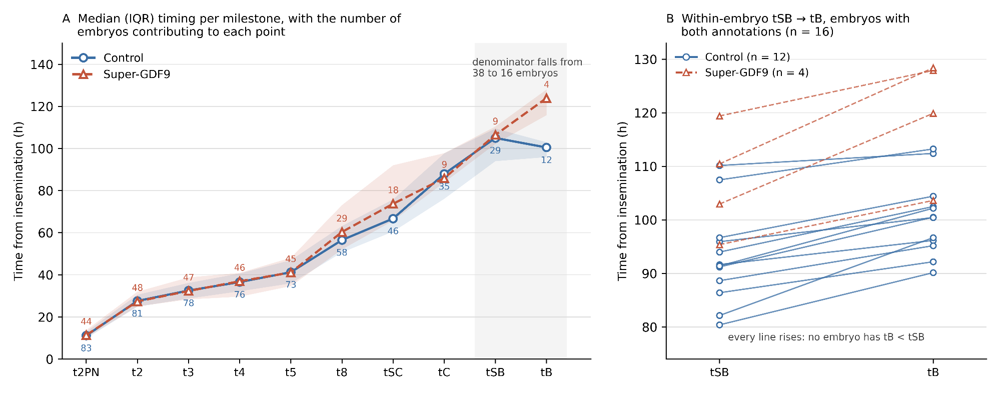
Longitudinal developmental progression and blastulation kinetics of human embryos cultured with or without super-GDF9 supplementation. Preimplantation developmental milestones from pronuclear appearance (t2PN) to full blastocyst expansion (tB) were recorded by time-lapse monitoring following CAPA-IVM culture in the absence (Control, blue circles) or presence of 50 ng/mL super-GDF9 (Super-GDF9, orange triangles). (A) Overall developmental trajectories showed parallel progression between groups from t2PN through compaction (tC) and start of blastulation (tSB). However, time to full blastocyst formation (tB) was significantly prolonged in the super-GDF9 group compared with controls (unadjusted mean ± SD: 120.0 ± 11.6 h vs. 100.5 ± 7.2 h, p = 0.036; linear mixed-effects model adjusted mean difference: 18.98 h, 95% CI: 8.63–29.34, p = 0.002). The apparent decrease in median timing between tSB and tB in the control arm reflects cohort attrition and the reduction in sample size from 29 embryos at tSB to 12 embryos at tB. (B) Paired trajectory analysis of individual embryos reaching the blastocyst stage (n = 16: 12 control, 4 super-GDF9) confirming that within every annotated embryo, blastulation progressed monotonically from tSB to tB without reversal. In Panel A, markers represent median timings (hours post-insemination) and shaded bands indicate the interquartile range (IQR); numbers adjacent to each data point indicate the number of embryos contributing to that milestone (shaded gray box highlights the attrition period from tSB to tB). In Panel B, connected lines represent paired trajectories for individual embryos. CAPA-IVM, capacitation in vitro maturation; sGDF9, super growth differentiation factor 9; t2PN, time to appearance of two pronuclei; t2–t8, time to 2- through 8-cell stages; tSC, time to start of compaction; tC, time to full compaction; tSB, time to start of blastulation; tB, time to full blastocyst development.
Morphokinetic data for later developmental milestones (tSC, tC, tSB, and tB) were available in a smaller cohort due to physiological embryo arrest and elective Day 3 interventions (Table 1). While timings for compaction onset (tSC: 68.8 ± 11.4 vs. 76.2 ± 18.1 h; p = 0.121), full compaction (tC: 86.1 ± 14.0 vs. 92.5 ± 13.7 h; p = 0.232), and initiation of blastulation (tSB: 102.1 ± 11.6 vs. 106.2 ± 8.1 h; p = 0.256) were not significantly different, the time to full blastocyst expansion (tB) was significantly prolonged in the sGDF9 group compared with controls (120.0 ± 11.6 vs. 100.5 ± 7.2 h; p = 0.036; analyzed in n = 16 blastocysts: 12 control and 4 sGDF9).
Multivariable Mixed-Effects Model Analysis
After accounting for patient-level clustering using linear mixed-effects models (Table 2), early cleavage parameters remained statistically comparable between groups (all unadjusted and BH-adjusted p > 0.05). Embryos cultured with sGDF9 demonstrated a non-significant trend toward delayed division to the 8-cell stage (t8: adjusted mean difference: 5.25 h; 95% CI: −0.45 to 10.94; nominal p = 0.070; BH-adjusted p = 0.387). Time to full blastocyst expansion remained significantly longer in the sGDF9 group (tB: adjusted mean difference: 18.98 h; 95% CI: 8.63–29.34; nominal p = 0.002; BH-adjusted p = 0.030).
Multivariable mixed-effects model analysis of embryo morphokinetic outcomes accounting for patient-level clustering.
| Outcome Parameter | Model Estimate | 95% Confidence Interval | Degrees of Freedom (df) | Nominal p-value | Embryos (Patients) | BH-Adjusted p-value |
|---|---|---|---|---|---|---|
| tSC (h) | 6.33 | −0.91, 13.57 | 60.7 | 0.086 | 64 (8) | 0.387 |
| tC (h) | 4.77 | −5.63, 15.16 | 42.0 | 0.360 | 44 (6) | 0.600 |
| tSB (h) | 2.22 | −5.97, 10.41 | 35.8 | 0.586 | 38 (7) | 0.749 |
| tB (h) | 18.98 | 8.63, 29.34 | 13.6 | 0.002 | 16 (7) | 0.030 |
| t8 (h) | 5.25 | −0.45, 10.94 | 83.1 | 0.070 | 87 (9) | 0.387 |
| s2 (t4 − t3) (h) | −1.33 | −2.97, 0.31 | 118.9 | 0.110 | 121 (9) | 0.387 |
| Direct cleavage 1 | 1.81* | 0.84, 3.88 | — | 0.129 | 135 (9) | 0.387 |
| s3 (t8 − t5) (h) | 3.80 | −1.56, 9.16 | 84.3 | 0.162 | 87 (9)† | 0.405 |
| Cell cycle 2 (t3 − t2) (h) | −1.52 | −4.14, 1.11 | 122.1 | 0.255 | 125 (9) | 0.546 |
| t2 (h) | 1.45 | −1.67, 4.57 | 126.2 | 0.360 | 129 (9) | 0.600 |
| t4 (h) | −1.35 | −5.10, 2.40 | 120.0 | 0.477 | 122 (9) | 0.715 |
| Direct cleavage 2 | 0.71* | 0.20, 2.52 | — | 0.599 | 135 (9) | 0.749 |
| t3 (h) | −0.63 | −4.43, 3.17 | 123.0 | 0.743 | 125 (9) | 0.857 |
| t5 (h) | −0.48 | −4.66, 3.69 | 115.8 | 0.820 | 118 (9) | 0.879 |
| t2PN (h) | 0.06 | −0.99, 1.11 | 120.7 | 0.911 | 127 (9) | 0.911 |
Abnormal Cleavage Patterns
The incidence of abnormal cleavage patterns is summarized in Table 1. Direct cleavage 1 (dc1) was the most prevalent abnormality observed in both cohorts, occurring in 45.9% (39/85) of control embryos and 62.0% (31/50) of sGDF9 embryos (unadjusted p = 0.077). In generalized linear mixed-effects analysis adjusting for patient clustering (Table 2), the odds of direct cleavage 1 were not significantly increased in the sGDF9 group compared with controls (conditional OR: 1.81; 95% CI: 0.84–3.88; nominal p = 0.129; BH-adjusted p = 0.387). Direct cleavage 2 was observed in 11.8% of control and 8.0% of sGDF9 embryos (conditional OR: 0.71; 95% CI: 0.20–2.52; p = 0.599). Reverse cleavage was rare, observed in 0% (0/85) of control embryos and 2.0% (1/50) of sGDF9 embryos (p = 0.370). Overall distributions of abnormal cleavage patterns did not differ significantly between the two groups (Fisher’s exact test, p = 0.117).
Discussion
In this secondary analysis of a prospective sibling-oocyte cohort, supplementation of the biphasic CAPA-IVM culture system with 50 ng/mL sGDF9 did not significantly alter early embryo cleavage kinetics or the incidence of cleavage abnormalities after adjusting for patient-level clustering using mixed-effects models. Nevertheless, notable kinetic trends were observed, specifically a non-significant delay in cleavage to the 8-cell stage and a statistically significant prolongation in the time required to achieve full blastocyst expansion (tB).
The substantial overlap in early cleavage timing distributions (t2PN through t8) between the control and sGDF9 groups indicates that sGDF9 supplementation during oocyte maturation does not substantially perturb early preimplantation mitotic divisions. This observation aligns with the established physiological role of GDF9, a member of the transforming growth factor-beta (TGF-β) superfamily, which functions primarily during pre-fertilization folliculogenesis and oocyte–cumulus cell communication rather than directly orchestrating post-fertilization cleavage events.24 Prior investigations have demonstrated the essential contribution of GDF9 to oocyte meiotic resumption and developmental competence, although evidence regarding its direct impact on post-cleavage embryogenesis has remained conflicting.25 A compelling mechanistic explanation for the lack of benefit observed here lies in signaling pathway selectivity: sGDF9 exclusively activates the SMAD2/3 signaling cascade, whereas physiological oocyte paracrine signaling relies on the synergistic actions of GDF9 and BMP15 (natively represented as cumulin), which concurrently stimulate both the SMAD2/3 and SMAD1/5/8 pathways.14 Balanced, dual-pathway activation is critical for orchestrating normal cumulus cell metabolism, extracellular matrix expansion, and steroidogenesis. The isolated activation of SMAD2/3 by sGDF9 in the absence of complementary BMP15 signaling may generate an incomplete or unbalanced signaling state, thereby limiting its capacity to enhance downstream embryonic competence.
Although not reaching statistical significance after multi-testing correction, embryos in the sGDF9 group exhibited an adjusted 5.25-hour delay in reaching the 8-cell stage (t8; p = 0.070), alongside a significant 18.98-hour delay in achieving full blastocyst expansion (tB; p = 0.002). In mammalian embryogenesis, the transition to the 8-cell stage represents the critical window of major embryonic genome activation (EGA), during which developmental control shifts from maternal messenger RNA and protein reserves to de novo embryonic transcription.26,27 Disruptions or delays during EGA are known to impair developmental progression, compromise blastocyst cell allocation, and attenuate implantation competence.27 Morphokinetic investigations have demonstrated that even minor kinetic deviations at key developmental junctions can reflect underlying metabolic or transcriptional stress.28 The delayed blastulation observed in the sGDF9 cohort may indicate that supplementation did not adequately facilitate the accumulation of essential maternal transcripts and metabolic substrates during oocyte maturation required to sustain robust EGA. However, this interpretation must be framed as an exploratory hypothesis: the blastocyst cohort was modest (n = 16 blastocysts), and the adjusted difference at t8 did not reach statistical significance. Blastulation kinetics are inherently multifactorial, governed by embryo intrinsic genetics, microenvironmental culture conditions, and epigenetic remodeling.24,29 Adequately powered studies are required to establish whether this kinetic lag has lasting functional consequences.
Regarding mitotic fidelity, unadjusted analysis revealed a higher frequency of direct cleavage 1 (dc1) in the sGDF9 group (62.0% vs. 45.9%), although this difference was not statistically significant in multivariable mixed-effects modeling (conditional OR: 1.81; 95% CI: 0.84–3.88; p = 0.129). Direct unequal cleavage, particularly during the first mitotic division from 1 to 3 blastomeres, is strongly linked to multipolar spindle formation, chromosome mis-segregation, embryonic aneuploidy, and diminished implantation rates.30 While the elevated incidence of dc1 in the sGDF9 group raises questions regarding potential mitotic destabilization, the lack of statistical significance after accounting for patient clustering indicates that patient-specific variation and baseline cohort characteristics likely account for this observation.
An intriguing insight from our findings is the contrast between single-phase IVM and biphasic CAPA-IVM systems regarding responsiveness to OSF supplementation. In preclinical mouse models, supplementing standard one-step IVM media with sGDF9 or cumulin consistently enhanced cumulus cell mucification, stimulated glycolytic gene expression, and increased blastocyst formation rates.8,24 Conversely, adding sGDF9 during biphasic CAPA-IVM failed to improve meiotic maturation, cleavage, or blastocyst yield in murine experiments, despite inducing transcriptional upregulation of cumulus expansion markers such as Ptgs2, Ptx3, and Adamts1.9,21 This discrepancy likely stems from the specialized biochemical architecture of CAPA-IVM. In CAPA-IVM, the pre-IVM phase employs cAMP modulators to maintain meiotic arrest and preserve transzonal projections, already optimizing cumulus–oocyte metabolic exchange and mitochondrial function.11 Under these optimized conditions, exogenous sGDF9 supplementation may provide redundant stimulation or perturb the fine-tuned metabolic balance achieved during the capacitation phase. Consequently, current evidence suggests that while sGDF9 holds clear promise for optimizing conventional, uncoordinated IVM protocols, its integration into biphasic CAPA-IVM systems requires careful reassessment of dosing, timing, and companion factors.
A principal methodological strength of this study is the application of linear and generalized linear mixed-effects models with patient-level random intercepts. Because human sibling oocytes and embryos derived from the same patient share intrinsic genetic and clinical backgrounds, failing to account for intra-cluster correlation can artificially inflate type I error rates.31 By modeling patient-level clustering, we ensured robust estimation of effect sizes. Furthermore, continuous time-lapse incubation enabled objective, fine-grained temporal tracking under undisturbed culture conditions.
Nevertheless, several limitations warrant consideration. First, as a secondary analysis of a clinical trial originally powered to evaluate oocyte maturation rates rather than embryo morphokinetics, the study had limited statistical power to detect small effect sizes in kinetic intervals. Second, inevitable attrition across extended preimplantation culture markedly reduced sample size at advanced developmental stages, with only 16 embryos (12 control and 4 sGDF9 across seven patients) reaching the fully expanded blastocyst stage. This late-stage attrition compromised the balanced sibling pairing present at earlier stages, meaning that blastocyst-stage kinetic findings must be interpreted strictly as exploratory and conditional upon blastocyst attainment. Finally, preimplantation genetic testing for aneuploidies (PGT-A) was not performed on these research embryos, precluding correlation between observed morphokinetic deviations and chromosomal ploidy.
Conclusion
In conclusion, supplementation of the biphasic CAPA-IVM culture system with 50 ng/mL sGDF9 was not associated with significant alterations in early human embryo cleavage kinetics or the incidence of abnormal cleavage patterns after adjusting for patient-level clustering. Although time to full blastocyst expansion was significantly prolonged in the sGDF9 group, this finding was based on a small subset of blastocysts and should be considered hypothesis-generating. Taken together, these data suggest that the pro-developmental effects of sGDF9 demonstrated in conventional single-step IVM do not readily translate into improved embryogenesis within biphasic CAPA-IVM protocols. Larger, prospectively powered trials evaluating balanced OSF combinations (such as cumulin) across titrated concentrations and incorporating genetic outcomes will be essential to establish the therapeutic utility of growth factor modulation in assisted reproductive technology.
Declarations
Abbreviations
AMH: anti-Müllerian hormone; ASRM: American Society for Reproductive Medicine; BH: Benjamini–Hochberg; BMI: body mass index; BMP15: bone morphogenetic protein 15; cAMP: cyclic adenosine monophosphate; CAPA-IVM: capacitation in vitro maturation; CI: confidence interval; COCs: cumulus–oocyte complexes; dc1: direct cleavage 1; dc2: direct cleavage 2; df: degrees of freedom; EGA: embryonic genome activation; ESHRE: European Society of Human Reproduction and Embryology; GDF9: growth differentiation factor 9; ICH-GCP: International Council for Harmonisation–Good Clinical Practice; ICSI: intracytoplasmic sperm injection; IQR: interquartile range; IVF: in vitro fertilization; IVM: in vitro maturation; OHSS: ovarian hyperstimulation syndrome; OR: odds ratio; OSFs: oocyte-secreted factors; PCOS: polycystic ovary syndrome; PGT-A: preimplantation genetic testing for aneuploidies; PMOS: polyendocrine metabolic ovarian syndrome; rc: reverse cleavage; REML: restricted maximum likelihood; s2: synchronicity of the second cell cycle; s3: synchronicity of the third cell cycle; SD: standard deviation; sGDF9: super growth differentiation factor 9; t2–t8: time to cleavage divisions from 2- to 8-cell stages; t2PN: time to appearance of two pronuclei; tB: time to full blastocyst development; tC/tM: time to compaction/morula; TGF-β: transforming growth factor-beta; tSB: time to start of blastulation; tSC: time to start of compaction.
Acknowledgments
We sincerely thank Associate Professor Craig A. Harrison and Dr. William A. Stocker of Monash University (Melbourne, Australia) for their generous provision of the Super-GDF9 used in this study. We also express our gratitude to the clinical, embryological, and laboratory staff of My Duc Hospital for their institutional support and assistance throughout this research.
Author’s contributions
All authors contributed to the conceptualization and design of the study and oversaw data collection. Data analysis was performed by TDP. The initial manuscript draft was planned and written by KTH, VNAH, AHL, CTT, TDP, MHNM, and NHT. LNV contributed to the critical revision of the manuscript. All authors participated in data interpretation, critically revised the manuscript for important intellectual content, and approved the final manuscript for publication.
Funding
This study was supported by a research grant from the Vietnam National Foundation for Science and Technology Development (NAFOSTED) under grant number FWO.108-2022.01.
Availability of data and materials
All processed data generated or analyzed during this study are fully presented within this manuscript. De-identified individual participant data and raw time-lapse annotations underlying this article will be shared upon reasonable request to the corresponding author.
Ethics approval and consent to participate
This study is a secondary analysis of a prospective sibling-oocyte cohort study conducted in Ho Chi Minh City, Vietnam (ClinicalTrials.gov identifier: NCT06766604). The study protocol was reviewed and approved by the Institutional Ethics Committee of My Duc Hospital, Ho Chi Minh City, Vietnam (approval reference: 12/24/DD-BVMD; approved on November 25, 2024). All participants received detailed counseling regarding study procedures, objectives, potential benefits, and risks, and provided written informed consent prior to enrollment.
Consent for publication
None (not applicable; no individual patient identifiers, images, or personal details are reported).
Declaration of generative AI and AI-assisted technologies in the writing process
During the preparation of this work, the authors used ChatGPT (OpenAI) to assist with English language editing and improving clarity of expression. Following this assistance, the authors reviewed and edited the content as needed and take full responsibility for the integrity and accuracy of the published article.
Competing interests
TMH has received speaker fees and honoraria from Merck and Ferring Pharmaceuticals. All other authors declare that they have no competing financial interests, commercial affiliations, or personal relationships that could have influenced the work reported in this paper.