


Association Between Acrosome-Related Sperm Head Abnormalities and Embryo Developmental Competence in First Intracytoplasmic Sperm Injection Cycles: A Retrospective Cohort Study
- Assisted Reproductive Technology Department, An Sinh Hospital, Ho Chi Minh City, Vietnam
Abstract
Introduction: Acrosome-related sperm head abnormalities are frequently encountered during intracytoplasmic sperm injection (ICSI); however, their independent influence on subsequent embryo developmental competence remains uncertain. This study investigated the association between acrosome-related sperm head abnormalities and embryological outcomes in first ICSI cycles after minimizing major female-related confounding factors.
Methods: This single-center retrospective cohort study analyzed 742 consecutive first ICSI cycles performed between January 2024 and December 2025 in women aged <35 years without severe oocyte dysmorphisms. The cohort comprised 370 cycles exclusively utilizing spermatozoa with acrosome-related head abnormalities (acrosomal area occupying <20% of the sperm head) and 372 control cycles utilizing spermatozoa with normal head morphology. The predefined primary outcome was the blastulation rate per injected mature (metaphase II [MII]) oocyte. Multivariable grouped quasi-binomial logistic regression and negative binomial regression models were fitted to adjust for maternal age, maternal body mass index (BMI), serum anti-Müllerian hormone (AMH) concentration, and controlled ovarian stimulation protocol.
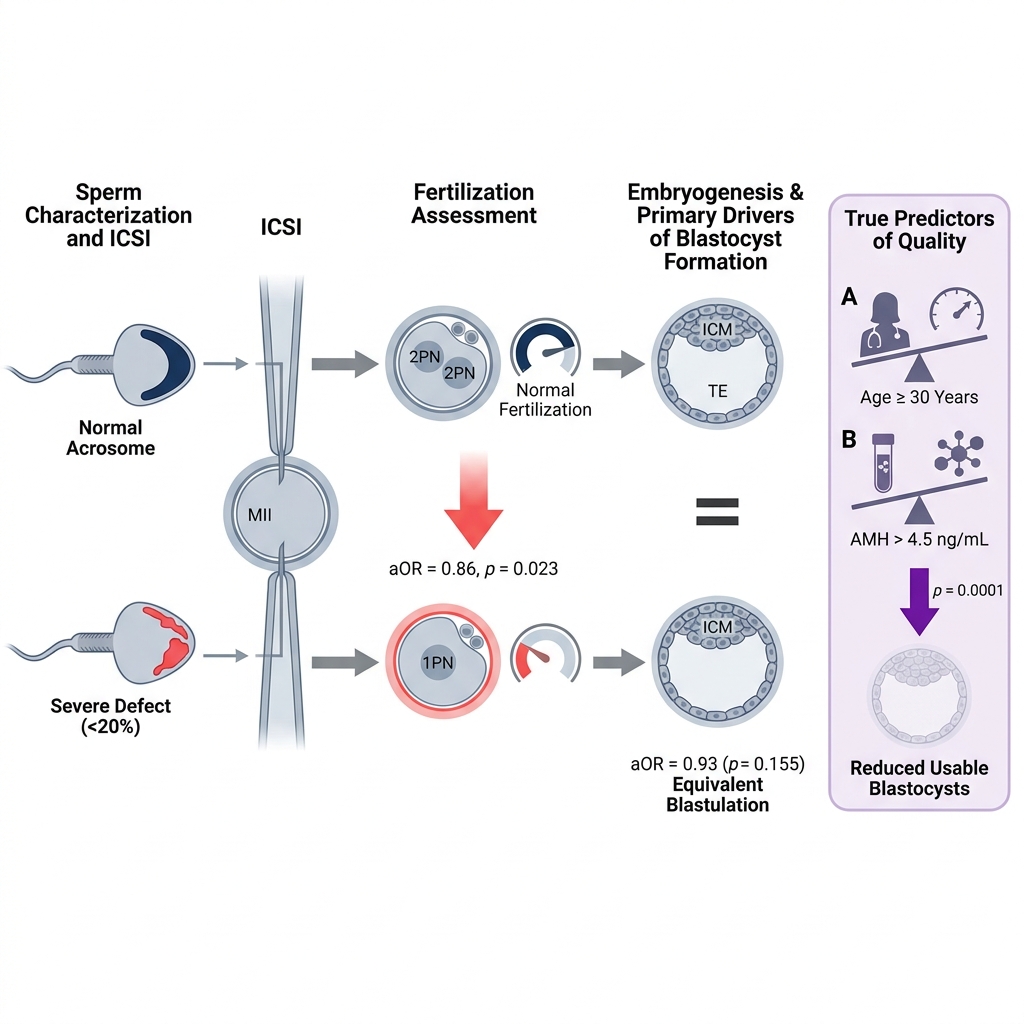
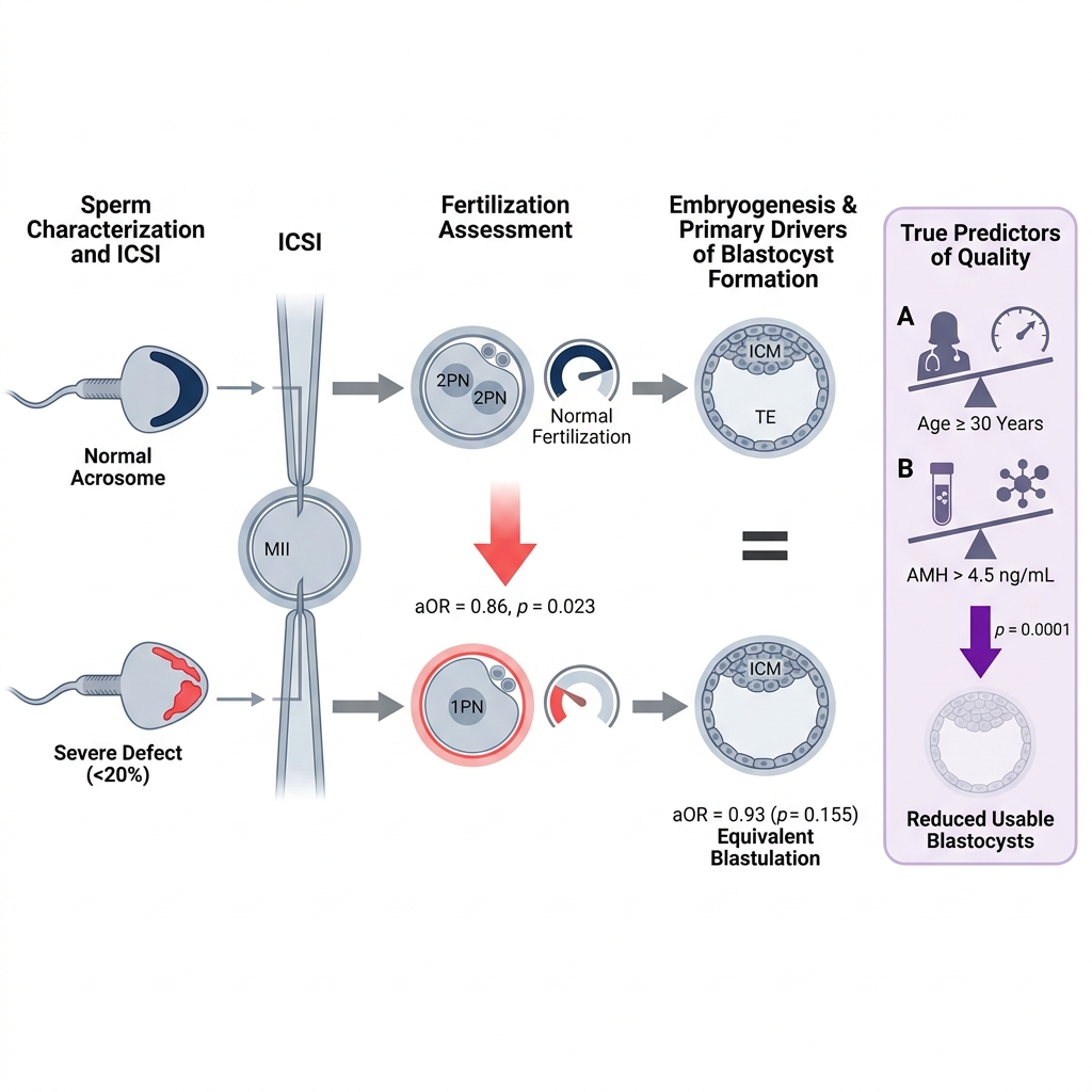
Results: The primary outcome, blastulation rate, did not differ significantly between the acrosome-related abnormalities group and the control group (median 48% vs. 50%; P = 0.1775). In unadjusted comparisons, cycles utilizing spermatozoa with acrosome-related abnormalities demonstrated significantly lower fertilization rates (84% vs. 88%; P = 0.0085), good-quality cleavage-stage embryo rates (58% vs. 61%; P = 0.0332), and good-quality blastocyst rates (17% vs. 20%; P = 0.0480). After multivariable adjustment, acrosome-related sperm head abnormalities remained independently associated with a reduced fertilization rate (adjusted odds ratio [aOR] 0.86, 95% confidence interval [CI] 0.76–0.98; P = 0.023). Conversely, adjusted associations with the blastulation rate (aOR 0.93, 95% CI 0.83–1.03; P = 0.155), usable blastocyst rate (aOR 0.91, 95% CI 0.82–1.01; P = 0.077), and total number of usable blastocysts per cycle (adjusted incidence rate ratio [aIRR] 0.94, 95% CI 0.86–1.01; P = 0.106) were not statistically significant. Exploratory interaction analyses revealed no significant effect modification by maternal age, ovarian reserve, number of MII oocytes, or ovarian stimulation protocol (all P for interaction > 0.05).
Conclusion: Acrosome-related sperm head abnormalities were independently associated with a lower fertilization rate in first ICSI cycles after controlling for key female-related factors, whereas multivariable-adjusted associations with blastulation and usable blastocyst rates did not reach statistical significance. While acrosomal defects compromise oocyte fertilization following ICSI, their independent impact on subsequent preimplantation embryo developmental competence appears limited.
Graphical abstract
Introduction
Intracytoplasmic sperm injection (ICSI) has revolutionized the management of male-factor infertility by bypassing the physiological barriers that impede natural sperm–oocyte interaction1. Nevertheless, fertilization and preimplantation embryo development rates remain variable following ICSI, indicating that intrinsic paternal factors continue to influence reproductive outcomes beyond the mechanical delivery of a spermatozoon into the ooplasm. Among the structural components of the sperm head, the acrosome plays an essential role in physiological fertilization and is closely linked to nuclear remodeling and functional maturation during spermiogenesis2,3. Although ICSI circumvents the requirement for acrosome-mediated penetration of the zona pellucida and cumulus oophorus, morphological defects of the acrosome frequently coexist with underlying ultrastructural or biochemical deficiencies that remain relevant to oocyte activation and subsequent embryonic development.
The prognostic value of conventional sperm morphology for assisted reproductive technology (ART) outcomes remains debated. Whereas routine semen morphology assessments have demonstrated limited ability to predict blastocyst formation following ICSI4,5, growing evidence suggests that targeted evaluation of specific sperm head defects at high magnification provides superior prognostic insight. Structural abnormalities involving the sperm neck, nucleus, and acrosome have been associated with impaired fertilization, arrested embryo cleavage, and reduced blastulation6,7. Acrosome-related abnormalities are of particular clinical and biological interest because aberrant acrosome biogenesis may coincide with defective sperm maturation, oocyte activation deficiency, impaired chromatin condensation, and compromised genomic integrity8,9,10. Specifically, reduced fertilization rates have been documented in men presenting with partial acrosomal hypoplasia, complete absence of the acrosome (globozoospermia), and related acrosomal disorders11,12,13. However, whether acrosome-related sperm head defects independently compromise post-fertilization embryo developmental competence following ICSI remains unclear.
Interpreting the existing literature is further complicated by the substantial contribution of oocyte quality to early embryogenesis. Maternal age, ovarian reserve, ovarian response to stimulation, and cytoplasmic morphology are primary determinants of fertilization, embryonic genome activation, and blastocyst formation, and high-competence oocytes may partially repair or compensate for paternal defects during early cleavage stages9,14. Most prior studies have enrolled heterogeneous female cohorts or focused predominantly on fertilization outcomes, making it difficult to isolate the intrinsic contribution of acrosome-related sperm abnormalities from variability in oocyte developmental competence. To minimize female-related confounding, the present study restricted analysis to first ICSI cycles in women aged <35 years with favorable ovarian response and excluded cycles exhibiting severe oocyte dysmorphisms. Accordingly, we aimed to investigate whether acrosome-related sperm head abnormalities are independently associated with embryological outcomes in first ICSI cycles—with the blastulation rate per injected mature oocyte designated as the predefined primary outcome—after minimizing heterogeneity in key female reproductive characteristics.
Materials and Methods
Study design and participants
This single-center, retrospective cohort study included consecutive couples undergoing their first ICSI cycle between January 2024 and December 2025 at the Assisted Reproductive Technology Department, An Sinh Hospital (Ho Chi Minh City, Vietnam). In routine clinical practice at our center, ICSI is widely performed as the standard fertilization method during in vitro fertilization (IVF) treatment, including in first cycles without severe oligozoospermia or asthenozoospermia. The clinical decision to perform ICSI was made by the attending physician prior to treatment initiation and was independent of this retrospective investigation. Only cycles utilizing fresh autologous oocytes with a planned indication for extended blastocyst culture were eligible for inclusion. Eligible female partners were younger than 35 years and had no documented medical or gynecological conditions expected to adversely affect ovarian response or oocyte quality.
All included cycles utilized freshly ejaculated autologous semen samples demonstrating normal sperm concentration (≥16 × 10 spermatozoa/mL) and progressive motility (≥30%) according to the World Health Organization (WHO) Laboratory Manual for the Examination and Processing of Human Semen (6th edition). Exposure status was defined at the treatment-cycle level rather than the individual spermatozoon level, as real-time sperm selection for ICSI reflects the morphological profile of the entire viable sperm population available at injection. The study group (acrosome-related abnormalities group) comprised cycles in which no morphologically normal spermatozoa could be identified and all spermatozoa selected for injection exhibited severe acrosome-related head abnormalities. The control group consisted of couples meeting identical maternal and seminal eligibility criteria who underwent ICSI using spermatozoa with normal head and acrosomal morphology.
Cycles utilizing cryopreserved sperm, donor sperm, or surgically retrieved sperm—including percutaneous epididymal sperm aspiration (PESA), conventional testicular sperm extraction (TESE), or microdissection TESE (micro-TESE)—were excluded. Additional exclusion criteria were rescue ICSI, predominant sperm morphological defects other than acrosome-related head abnormalities, severe intracytoplasmic or extracytoplasmic oocyte dysmorphisms known to compromise embryo developmental competence (specifically large or multiple cytoplasmic vacuoles, centrally located cytoplasmic granulation, or smooth endoplasmic reticulum [SER] aggregates), and cycles yielding fewer than six mature (metaphase II [MII]) oocytes for injection. The lower cutoff of six MII oocytes was predefined to reduce variance attributable to suboptimal ovarian response, as prior evidence indicates that cycles retrieving approximately 6–15 mature oocytes exhibit more uniform oocyte developmental competence than low-yield cycles15. Each couple contributed only their first treatment cycle to eliminate repeated-measures bias.
The minimum required sample size was calculated using the standard formula for comparing two independent proportions. Based on the historical blastulation rate at our center (52%) and prior evidence linking severe sperm head teratozoospermia to reduced blastocyst formation16, an expected blastulation rate of 40% was assumed for the acrosome-related abnormalities group. Assuming a two-sided significance level (α) of 0.05 and 80% statistical power (1 − β = 0.80), a minimum of 270 treatment cycles per group was required.
Controlled ovarian stimulation and ICSI procedures
Controlled ovarian stimulation (COS) was conducted using either a progestin-primed ovarian stimulation (PPOS) protocol or a gonadotropin-releasing hormone (GnRH) antagonist protocol. In routine clinical practice at our center, the attending clinician selected the stimulation regimen based on the planned embryo transfer strategy: the PPOS protocol was preferentially employed when a freeze-all strategy was planned, whereas the GnRH antagonist protocol was selected when fresh embryo transfer was anticipated.
In the PPOS regimen, ovarian stimulation was performed using highly purified menotropin (IVF-M, LG Chem Ltd., Seoul, Republic of Korea) co-administered with oral dydrogesterone (Duphaston, Abbott Biologicals B.V., Olst, the Netherlands; 10 mg twice daily) from the initiation of gonadotropin stimulation until the day of the final oocyte maturation trigger. In the GnRH antagonist regimen, ovarian stimulation was performed using menotropin (IVF-M) with flexible introduction of cetrorelix acetate (Cetrotide, Merck Europe B.V., Amsterdam, the Netherlands). Final oocyte maturation was induced using a dual trigger (triptorelin plus human chorionic gonadotropin) once at least three leading follicles reached a mean diameter of ≥17 mm, followed by transvaginal ultrasound-guided oocyte retrieval 36–38 hours later.
During routine clinical practice, real-time sperm selection for ICSI was performed by senior embryologists under an inverted microscope at ×600 magnification. All embryologists performing ICSI were trained under a single standardized laboratory protocol and applied uniform morphological selection criteria throughout the study period. Morphologically normal spermatozoa were preferentially selected whenever present. Acrosomal morphology was evaluated by estimating the proportion of the anterior sperm head occupied by the acrosomal region. A cycle was assigned to the acrosome-related abnormalities group only when no morphologically normal spermatozoa could be identified in the processed sample and every spermatozoon selected for ICSI exhibited an acrosomal area occupying <20% of the sperm head (including occasional spermatozoa with complete absence of the acrosome); no cases of complete (100%) globozoospermia were present in the analytic cohort. This <20% threshold was predefined because it represents a marked departure from the physiological acrosomal coverage of 40%–70% of the sperm head specified in the WHO Laboratory Manual (6th edition), thereby ensuring clear morphological separation between the exposure and control groups. This conservative institutional criterion was established by consensus among senior embryologists at our center to maximize consistency and minimize subjective misclassification during real-time assessment of unstained, motile spermatozoa. Spermatozoa displaying other major head defects (e.g., macrocephaly, pinhead, or multiple heads) were deemed unsuitable for clinical injection and were excluded from both clinical use and the present analysis.
ICSI was performed on all MII oocytes, and injected oocytes were subsequently cultured in Continuous Single Culture Medium (CSCM; FUJIFILM Irvine Scientific, Santa Ana, CA, USA) through to the blastocyst stage.
Embryo culture and morphological assessment
Fertilization was evaluated 16–18 hours post-ICSI; normal fertilization was defined by the presence of two distinct pronuclei (2PN) and two polar bodies. Because embryos were cultured in a microdrop group-culture system, individual zygotes could not be tracked longitudinally to the blastocyst stage; consequently, pronuclear morphology (Z-score grading) was not analyzed.
Cleavage-stage embryos were evaluated 67–69 hours post-ICSI in accordance with the Alpha Scientists in Reproductive Medicine and ESHRE Istanbul Consensus criteria. Blastocyst morphology was graded 115–117 hours post-ICSI using the Gardner and Schoolcraft grading system. Good-quality cleavage-stage embryos were defined as grade 1 or grade 2 embryos with stage-appropriate blastomere number and <20% fragmentation. Good-quality blastocysts were defined as expanded or hatching blastocysts (expansion grade ≥3) with an inner cell mass (ICM) and trophectoderm (TE) graded as AA, AB, BA, or BB (grade 1 or grade 2 institutional equivalent). Usable blastocysts were defined as all blastocysts meeting institutional clinical criteria for either fresh embryo transfer or cryopreservation. In cycles utilizing the PPOS protocol, all usable blastocysts were vitrified. In cycles utilizing the GnRH antagonist protocol, up to two embryos were selected for fresh transfer, and all supernumerary usable blastocysts were vitrified using the Cryotech Vitrification Kit 101 (Cryotech, Tokyo, Japan).
Baseline variables
Baseline clinical and laboratory covariates extracted from electronic medical records included maternal age, paternal age, maternal body mass index (BMI), baseline serum anti-Müllerian hormone (AMH) concentration, COS protocol (PPOS vs. GnRH antagonist), duration of ovarian stimulation (days), total follicle-stimulating hormone (FSH)/menotropin dose administered (IU), total number of retrieved cumulus–oocyte complexes, number of mature (MII) oocytes, ejaculated sperm concentration (×10^6/mL), and progressive sperm motility (%).
Outcome measures
The predefined primary outcome was the blastulation rate, calculated as the proportion of injected MII oocytes that developed to the blastocyst stage. Predefined secondary outcomes included the normal fertilization rate, cleavage-stage embryo rate, good-quality cleavage-stage embryo rate, good-quality blastocyst rate, usable blastocyst rate, and the absolute number of usable blastocysts generated per treatment cycle.
Specifically, the fertilization rate was defined as the number of 2PN zygotes divided by the total number of injected MII oocytes. The cleavage-stage embryo rate and good-quality cleavage-stage embryo rate were defined as the proportions of injected MII oocytes developing into cleavage-stage embryos and good-quality cleavage-stage embryos on Day 3, respectively. Similarly, the good-quality blastocyst rate and usable blastocyst rate were defined as the proportions of injected MII oocytes developing into good-quality blastocysts and clinically usable blastocysts on Day 5, respectively. All proportional embryological outcomes were computed at the patient (cycle) level using the number of injected MII oocytes as the denominator, irrespective of whether embryos were subsequently transferred fresh or vitrified. In addition, the total number of usable blastocysts obtained per cycle was analyzed as a discrete count outcome.
Statistical analysis
Continuous variables were evaluated for normality using the Shapiro–Wilk test. Normally distributed continuous variables are expressed as mean ± standard deviation (SD) and were compared using the independent-samples Student's t-test; non-normally distributed continuous variables are expressed as median (interquartile range [IQR]) and were compared using the Mann–Whitney U test. Categorical variables are presented as frequency (percentage) and were compared using Pearson's chi-square (χ2) test or Fisher's exact test, as appropriate.
To account for within-cycle clustering of oocytes and inter-patient variability in MII oocyte yield, multivariable generalized linear models (GLMs) with a quasi-binomial distribution and logit link function were fitted to estimate the independent associations between acrosome-related sperm head abnormalities and the fertilization rate, blastulation rate, and usable blastocyst rate. Outcomes were parameterized as grouped binomial counts (number of successes versus number of failures out of total injected MII oocytes per cycle), with each patient contributing a single independent observation. Multivariable models were adjusted for a priori selected female and treatment covariates: maternal age (<30 vs. ≥30 years), maternal BMI (≤23.0 vs. >23.0 kg/m2), serum AMH concentration (≤4.5 vs. >4.5 ng/mL), and COS protocol (GnRH antagonist vs. PPOS). Extra-binomial variation (overdispersion) was quantified using the Pearson dispersion parameter (φ) and accounted for via the quasi-binomial variance structure. Effect estimates are reported as adjusted odds ratios (aORs) with 95% confidence intervals (CIs). Dichotomous cutoffs for maternal age (<30 vs. ≥30 years), serum AMH concentration (≤4.5 vs. >4.5 ng/mL), and maternal BMI (≤23.0 vs. >23.0 kg/m2) were selected based on established clinical thresholds in reproductive medicine17,18 and the WHO Western Pacific/Asia-Pacific BMI classification for Asian populations.
To evaluate predictors of the absolute number of usable blastocysts yielded per treatment cycle, a multivariable negative binomial regression model with a log link function was fitted, adjusting for the same set of covariates; results are reported as adjusted incidence rate ratios (aIRRs) with 95% CIs.
Pre-specified exploratory interaction analyses were performed by incorporating multiplicative interaction terms into the quasi-binomial models to test whether the association between acrosome-related sperm head abnormalities and embryological outcomes varied across strata of maternal age (<30 vs. ≥30 years), ovarian reserve (AMH ≤4.5 vs. >4.5 ng/mL), ovarian response (number of MII oocytes, modeled continuously), or COS protocol (PPOS vs. GnRH antagonist).
All statistical analyses were performed using R software (version 4.4.2; R Foundation for Statistical Computing, Vienna, Austria). A two-sided P value < 0.05 was considered statistically significant.
Results
Study cohort and baseline characteristics
A total of 2,453 consecutive first ICSI cycles performed between January 2024 and December 2025 were screened for eligibility. Of these, 349 cycles were excluded because embryos were not cultured to the blastocyst stage, and 1,362 cycles were excluded based on the predefined clinical, seminal, and oocyte exclusion criteria. The final analytic cohort comprised 742 cycles, including 370 cycles in the acrosome-related abnormalities group and 372 cycles in the morphologically normal control group (Figure 1).
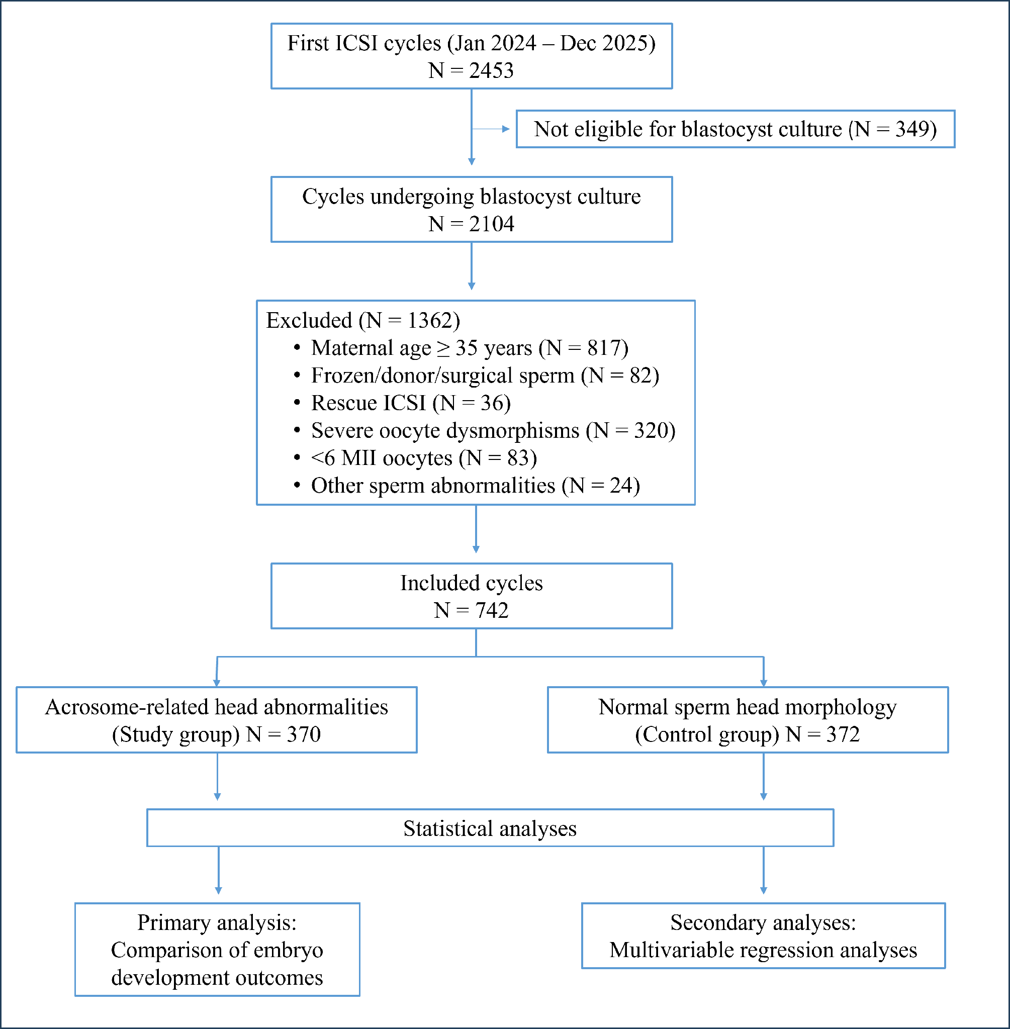
Patient screening, eligibility assessment, and cohort allocation flowchart for first intracytoplasmic sperm injection (ICSI) cycles performed at An Sinh Hospital between January 2024 and December 2025. A total of 2,453 consecutive first ICSI cycles in women aged <35 years were screened. Following the exclusion of cycles without blastocyst culture (n = 349) and cycles meeting predefined clinical, seminal, or oocyte exclusion criteria (n = 1,362; including frozen, donor, or surgically retrieved sperm [PESA, TESE, or micro-TESE], rescue ICSI, non-acrosomal sperm head defects, severe oocyte dysmorphisms, or <6 mature metaphase II [MII] oocytes retrieved), 742 eligible cycles were included in the final analysis. Eligible cycles were stratified into the acrosome-related sperm head abnormalities group (acrosomal area occupying <20% of the sperm head across all injected spermatozoa; n = 370) and the morphologically normal control group (normal sperm head and acrosomal morphology; n = 372). Abbreviations: ICSI, intracytoplasmic sperm injection; micro-TESE, microdissection testicular sperm extraction; MII, metaphase II; PESA, percutaneous epididymal sperm aspiration; TESE, testicular sperm extraction.
Baseline parental demographic characteristics, ovarian reserve markers, gonadotropin stimulation parameters, oocyte yield, and conventional semen parameters were well balanced between the two groups (Table 1). Specifically, no statistically significant differences were observed between the acrosome-related abnormalities group and the control group with respect to maternal age, paternal age, maternal BMI, serum AMH concentration, duration of ovarian stimulation, total gonadotropin dose, number of retrieved oocytes, number of injected MII oocytes, sperm concentration, or progressive sperm motility (all P > 0.05). The sole baseline difference between groups was the distribution of COS protocols: a higher proportion of cycles in the acrosome-related abnormalities group utilized the PPOS protocol (57% vs. 34%), whereas the GnRH antagonist protocol was more frequent in the control group (66% vs. 43%; P < 0.001) (Table 1).
Baseline demographic, ovarian stimulation, oocyte, and conventional semen characteristics of couples undergoing first ICSI cycles stratified by sperm acrosomal morphology (N = 742).
| Characteristic | Control Group (Normal Sperm Head Morphology, n = 372) | Study Group (Acrosome-Related Sperm Head Abnormalities, n = 370) | P value |
|---|---|---|---|
| Continuous variables, median (IQR) | |||
| Maternal age (years) | 31 (28–33) | 31 (28–34) | 0.09 |
| Paternal age (years) | 33 (30–37) | 34 (31–37) | 0.20 |
| Maternal BMI (kg/m²) | 21.84 (20.00–23.63) | 22.00 (20.05–23.83) | 0.80 |
| Serum AMH concentration (ng/mL) | 4.08 (2.64–6.07) | 3.94 (2.43–6.09) | 0.50 |
| Ovarian stimulation duration (days) | 8 (8–9) | 8 (8–9) | 0.50 |
| Total gonadotropin (FSH) dose (IU) | 2,400 (2,400–2,700) | 2,400 (2,400–2,700) | 0.80 |
| Retrieved oocytes per cycle (n) | 19 (13–25) | 18 (13–25) | 0.60 |
| Mature (MII) oocytes per cycle (n) | 15 (11–20) | 15 (11–20) | 0.80 |
| Sperm concentration (×10⁶/mL) | 35 (25–45) | 35 (25–45) | 0.90 |
| Progressive sperm motility (%) | 50 (39–63) | 50 (39–63) | 0.90 |
| Controlled ovarian stimulation protocol, n (%) | <0.001 | ||
| PPOS protocol | 125 (34%) | 212 (57%) | |
| GnRH antagonist protocol | 247 (66%) | 158 (43%) |
Unadjusted embryological outcomes
Unadjusted patient-level embryological outcomes are summarized in Table 2. The predefined primary outcome, blastulation rate per injected MII oocyte, did not differ significantly between the acrosome-related abnormalities group and the control group (median 48% [IQR 35%–60%] vs. 50% [IQR 36%–63%]; P = 0.1775).
Among the secondary outcomes, cycles in the acrosome-related abnormalities group exhibited a significantly lower normal fertilization rate (median 84% [IQR 74%–92%] vs. 88% [IQR 78%–93%]; P = 0.0085), good-quality cleavage-stage embryo rate (58% [IQR 46%–70%] vs. 61% [IQR 48%–75%]; P = 0.0332), and good-quality blastocyst rate (17% [IQR 8%–29%] vs. 20% [IQR 11%–31%]; P = 0.0480) compared with the control group. Conversely, no statistically significant between-group differences were detected in the overall cleavage-stage embryo rate (72% [IQR 62%–82%] vs. 74% [IQR 63%–85%]; P = 0.1471) or the usable blastocyst rate (38% [IQR 25%–50%] vs. 38% [IQR 27%–50%]; P = 0.1283) (Table 2).
Unadjusted comparison of patient-level fertilization and preimplantation embryo development rates per injected metaphase II oocyte between the acrosome-related abnormalities group and the morphologically normal control group (N = 742).
| Embryological Outcome | Control Group (Normal Sperm Head Morphology, n = 372) | Study Group (Acrosome-Related Sperm Head Abnormalities, n = 370) | P value |
|---|---|---|---|
| Primary outcome | |||
| Blastulation rate (%) | 50 (36–63) | 48 (35–60) | 0.1775 |
| Secondary outcomes | |||
| Normal fertilization (2PN) rate (%) | 88 (78–93) | 84 (74–92) | 0.0085 |
| Cleavage-stage embryo rate (%) | 74 (63–85) | 72 (62–82) | 0.1471 |
| Good-quality cleavage-stage embryo rate (%) | 61 (48–75) | 58 (46–70) | 0.0332 |
| Good-quality blastocyst rate (%) | 20 (11–31) | 17 (8–29) | 0.0480 |
| Usable blastocyst rate (%) | 38 (27–50) | 38 (25–50) | 0.1283 |
Multivariable quasi-binomial regression analyses
Multivariable grouped quasi-binomial logistic regression models were fitted to evaluate the independent associations between acrosome-related sperm head abnormalities and proportional embryological outcomes after adjusting for maternal age, maternal BMI, serum AMH concentration, and COS protocol.
For the primary outcome, acrosome-related sperm head abnormalities were not significantly associated with the blastulation rate after multivariable adjustment (aOR 0.93, 95% CI 0.83–1.03; P = 0.155) (Table 3a).
Multivariable grouped quasi-binomial logistic regression analysis of factors associated with the primary outcome (blastulation rate per injected metaphase II oocyte) in first ICSI cycles (N = 742).
| Variable / Category | Blastulation Rate aOR (95% CI) | P value |
|---|---|---|
| Sperm acrosome morphology | ||
| Normal (≥40%–70% head coverage) | 1.00 (Reference) | — |
| Abnormal (<20% head coverage) | 0.93 (0.83–1.03) | 0.155 |
| Maternal age (years) | ||
| <30 | 1.00 (Reference) | — |
| ≥30 | 0.93 (0.84–1.04) | 0.207 |
| Maternal BMI (kg/m²) | ||
| ≤23.0 | 1.00 (Reference) | — |
| >23.0 | 0.92 (0.82–1.03) | 0.130 |
| Serum AMH concentration (ng/mL) | ||
| ≤4.5 | 1.00 (Reference) | — |
| >4.5 | 1.01 (0.91–1.12) | 0.852 |
| Controlled ovarian stimulation protocol | ||
| GnRH antagonist | 1.00 (Reference) | — |
| PPOS | 1.06 (0.95–1.18) | 0.312 |
For the secondary proportional outcomes, acrosome-related sperm head abnormalities remained independently associated with a 14% lower odds of normal oocyte fertilization per injected MII oocyte (aOR 0.86, 95% CI 0.76–0.98; P = 0.023). By contrast, the multivariable-adjusted association between acrosome-related abnormalities and the usable blastocyst rate was attenuated and did not reach statistical significance (aOR 0.91, 95% CI 0.82–1.01; P = 0.077) (Table 3b). None of the adjusted female covariates (maternal age, BMI, AMH concentration, or COS protocol) showed a statistically significant association with fertilization rate, blastulation rate, or usable blastocyst rate in the grouped quasi-binomial models. The estimated Pearson dispersion parameters were φ = 1.68 for the fertilization rate, φ = 2.10 for the blastulation rate, and φ = 1.85 for the usable blastocyst rate, confirming moderate overdispersion that was appropriately accounted for by the quasi-binomial variance specification.
Multivariable grouped quasi-binomial logistic regression analyses of factors associated with secondary proportional outcomes (normal fertilization rate and usable blastocyst rate per injected metaphase II oocyte) in first ICSI cycles (N = 742).
| Variable / Category | Normal Fertilization Rate aOR (95% CI) | P value | Usable Blastocyst Rate aOR (95% CI) | P value |
|---|---|---|---|---|
| Sperm acrosome morphology | ||||
| Normal | 1.00 (Reference) | — | 1.00 (Reference) | — |
| Abnormal (<20% head coverage) | 0.86 (0.76–0.98) | 0.023 | 0.91 (0.82–1.01) | 0.077 |
| Maternal age (years) | ||||
| <30 | 1.00 (Reference) | — | 1.00 (Reference) | — |
| ≥30 | 0.92 (0.81–1.05) | 0.208 | 0.97 (0.87–1.07) | 0.514 |
| Maternal BMI (kg/m²) | ||||
| ≤23.0 | 1.00 (Reference) | — | 1.00 (Reference) | — |
| >23.0 | 1.02 (0.89–1.16) | 0.817 | 1.01 (0.91–1.12) | 0.842 |
| Serum AMH concentration (ng/mL) | ||||
| ≤4.5 | 1.00 (Reference) | — | 1.00 (Reference) | — |
| >4.5 | 1.10 (0.97–1.26) | 0.134 | 0.96 (0.86–1.06) | 0.421 |
| Controlled ovarian stimulation protocol | ||||
| GnRH antagonist | 1.00 (Reference) | — | 1.00 (Reference) | — |
| PPOS | 1.08 (0.95–1.22) | 0.270 | 1.03 (0.93–1.14) | 0.586 |
Multivariable negative binomial regression analysis of usable blastocyst count
Because proportional rates per MII oocyte do not directly capture the cumulative number of embryos available for clinical transfer or cryopreservation per cycle, a multivariable negative binomial regression model was fitted to evaluate the total count of usable blastocysts obtained per cycle (Table 4).
Multivariable negative binomial regression analysis of clinical and seminal factors associated with the absolute number of usable blastocysts obtained per treatment cycle in first ICSI cycles (N = 742).
| Variable / Category | Usable Blastocyst Count per Cycle aIRR (95% CI) | P value |
|---|---|---|
| Sperm acrosome morphology | ||
| Normal | 1.00 (Reference) | — |
| Abnormal (<20% head coverage) | 0.94 (0.86–1.01) | 0.106 |
| Maternal age (years) | ||
| <30 | 1.00 (Reference) | — |
| ≥30 | 0.87 (0.80–0.94) | <0.001 |
| Maternal BMI (kg/m²) | ||
| ≤23.0 | 1.00 (Reference) | — |
| >23.0 | 1.03 (0.94–1.12) | 0.542 |
| Serum AMH concentration (ng/mL) | ||
| ≤4.5 | 1.00 (Reference) | — |
| >4.5 | 0.92 (0.85–1.00) | 0.038 |
| Controlled ovarian stimulation protocol | ||
| GnRH antagonist | 1.00 (Reference) | — |
| PPOS | 0.97 (0.90–1.06) | 0.520 |
After adjustment for maternal age, maternal BMI, serum AMH concentration, and COS protocol, acrosome-related sperm head abnormalities were not independently associated with the number of usable blastocysts yielded per cycle (aIRR 0.94, 95% CI 0.86–1.01; P = 0.106). Conversely, maternal age ≥30 years (aIRR 0.87, 95% CI 0.80–0.94; P < 0.001) and baseline serum AMH concentration >4.5 ng/mL (aIRR 0.92, 95% CI 0.85–1.00; P = 0.038) were independently associated with a lower count of usable blastocysts per cycle. Maternal BMI and COS protocol were not significantly associated with usable blastocyst count (Table 4).
Subgroup interaction analyses
Exploratory interaction models demonstrated no statistically significant effect modification between acrosome-related sperm head abnormalities and maternal age (<30 vs. ≥30 years), baseline serum AMH concentration (≤4.5 vs. >4.5 ng/mL), ovarian response (number of MII oocytes), or COS protocol (PPOS vs. GnRH antagonist) for the fertilization rate, blastulation rate, or usable blastocyst rate (all P for interaction > 0.05; Table 5).
Exploratory subgroup interaction analyses evaluating effect modification by maternal age, oocyte yield, ovarian reserve, and ovarian stimulation protocol on the association between acrosome-related sperm head abnormalities and embryological outcomes (N = 742).
| Clinical Effect Modifier | P for Interaction (Normal Fertilization Rate) | P for Interaction (Blastulation Rate) | P for Interaction (Usable Blastocyst Rate) |
|---|---|---|---|
| Maternal age (<30 vs. ≥30 years) | 0.566 | 0.248 | 0.575 |
| Ovarian response (number of MII oocytes, continuous) | 0.393 | 0.134 | 0.294 |
| Serum AMH concentration (≤4.5 vs. >4.5 ng/mL) | 0.420 | 0.537 | 0.951 |
| Controlled ovarian stimulation protocol (PPOS vs. GnRH antagonist) | 0.623 | 0.680 | 0.487 |
Discussion
Principal findings
The principal finding of this retrospective cohort study is that severe acrosome-related sperm head abnormalities (<20% acrosomal coverage) were independently associated with a lower normal fertilization rate in first ICSI cycles after controlling for key female-related confounding variables. Although cycles utilizing spermatozoa with acrosome-related abnormalities also displayed lower unadjusted rates of good-quality cleavage-stage embryos and good-quality blastocysts, neither the overall cleavage-stage embryo rate nor the primary outcome—blastulation rate—differed significantly between groups. Furthermore, after multivariable adjustment, associations between acrosome-related abnormalities and the blastulation rate, usable blastocyst rate, and total number of usable blastocysts per cycle were not statistically significant. Collectively, these results indicate that while acrosome-related sperm head defects independently impair fertilization following ICSI, fertilized zygotes that successfully progress past pronuclear formation retain largely preserved competence to reach the blastocyst stage.
In the multivariable count model, maternal age ≥30 years was independently associated with a 13% reduction in the number of usable blastocysts per cycle (aIRR 0.87, 95% CI 0.80–0.94; P < 0.001), consistent with age-related declines in oocyte yield and quality even among women younger than 35 years. Notably, a baseline serum AMH concentration >4.5 ng/mL was also associated with a marginally lower number of usable blastocysts (aIRR 0.92, 95% CI 0.85–1.00; P = 0.038). This finding may reflect residual confounding by subclinical polycystic ovary morphology or reduced per-oocyte developmental competence in hyper-responders rather than a direct deleterious effect of elevated ovarian reserve, and should be interpreted cautiously given the retrospective design and the absence of MII oocyte count as an offset in the negative binomial model.
Blastulation versus morphological embryo quality following ICSI
An intriguing observation of our study is that overall blastulation rates were comparable between groups, whereas unadjusted rates of good-quality cleavage-stage embryos and good-quality blastocysts were modestly lower in cycles affected by acrosome-related sperm abnormalities. This divergence highlights that blastocyst cavitation and morphological grading capture distinct dimensions of preimplantation development: the blastulation rate reflects the capacity of an fertilized oocyte to undergo compaction, cavitation, and blastocoel expansion, whereas good-quality blastocyst metrics additionally require cohesive inner cell mass organization and uniform trophectoderm cellularity.
Nevertheless, these unadjusted differences in morphological embryo quality should be interpreted with caution. The multivariable-adjusted association between acrosome-related sperm abnormalities and the clinically actionable endpoint—the usable blastocyst rate—did not reach statistical significance (P = 0.077), and secondary quality comparisons were unadjusted for multiple testing. Consequently, our data do not support a definitive independent deleterious effect of acrosome-related sperm abnormalities on post-fertilization embryo developmental competence.
Comparison with previous studies
The independent reduction in fertilization rate observed in our cohort aligns closely with the established physiological role of the acrosome and perinuclear theca in sperm-borne oocyte activation. Prior clinical investigations have consistently documented compromised fertilization or total fertilization failure in men with partial acrosomal hypoplasia, small acrosomes, or globozoospermia, even when mechanical sperm delivery via ICSI is employed11,12,13,19. By restricting our study population to young women (<35 years) with normal ovarian response and morphologically normal oocytes, our study extends prior observations by confirming that this fertilization deficit persists independently of female-related factors.
Interestingly, the pattern observed in our unadjusted analyses—preserved overall blastulation accompanied by a modest reduction in high-grade blastocysts—parallels findings from recent studies of elevated sperm DNA fragmentation, wherein overall blastocyst formation remained largely unaffected while embryo morphokinetics and the yield of top-quality blastocysts were compromised [20, 21]. However, because our multivariable analyses showed no statistically significant reduction in either overall blastulation or usable blastocyst yield, any downstream paternal effect of acrosomal abnormalities after successful fertilization appears modest when high-quality young oocytes are utilized.
Biological plausibility and putative mechanisms
From a developmental biology perspective, subtle impairments in cleavage and blastocyst morphology have been attributed to the 'late paternal effect,' whereby paternal genomic or epigenetic lesions manifest after embryonic genome activation at the 4- to 8-cell transition21,22,23. Furthermore, high-competence oocytes from young women possess robust maternal mRNA transcripts and DNA repair machinery capable of correcting moderate paternal chromatin lesions during the first cleavage division14,22,23, which may explain why fertilized oocytes in our <35-year female cohort achieved near-normal blastulation rates.
With respect to fertilization impairment, acrosome biogenesis occurs concurrently with nuclear elongation and histone-to-protamine replacement during spermiogenesis. Structural defects in acrosomal vesicle biogenesis are closely linked to perturbations in the perinuclear theca, resulting in reduced expression or abnormal localization of phospholipase C zeta (PLCζ)—the primary physiological sperm factor responsible for inducing intracellular calcium (Ca2+) oscillations and metaphase-to-anaphase transition in the MII oocyte8,10,13,19. Additionally, aberrant acrosomal morphogenesis has been associated with incomplete protamination, loosely packaged chromatin, and elevated sperm DNA fragmentation10,13,19. Because our retrospective study did not directly assay PLCζ expression, chromatin protamination, or sperm DNA fragmentation index (DFI), these mechanistic pathways remain hypothetical and warrant verification in prospective translational studies.
Clinical implications
Our findings have practical implications for patient counseling and laboratory management in ART. When embryologists encounter ejaculates in which all available spermatozoa exhibit severe acrosomal reduction (<20% of the head area) during routine ICSI, clinicians can counsel couples that although a modest reduction in the normal fertilization rate is anticipated, the likelihood that fertilized oocytes will progress into clinically usable blastocysts remains reassuringly favorable, particularly in women aged <35 years with good ovarian reserve.
Moreover, the selective impairment in fertilization provides a clear biological rationale for future randomized controlled trials investigating whether targeted adjuvants—such as assisted oocyte activation (AOA) with calcium ionophores or advanced microfluidic/high-magnification sperm selection (IMSI)—can normalize fertilization rates and optimize cumulative blastocyst yield in patients with severe acrosomal abnormalities.
Strengths and limitations
A major strength of the present study is its rigorous eligibility design, which was specifically structured to minimize female-related confounding. By restricting the cohort to first ICSI cycles in women younger than 35 years with at least six MII oocytes and excluding cycles with severe cytoplasmic or extracytoplasmic oocyte dysmorphisms, we substantially reduced variability in maternal oocyte competence. Furthermore, we employed grouped quasi-binomial GLMs to account for within-patient clustering of oocytes and extra-binomial overdispersion, alongside negative binomial modeling for embryo counts and formal interaction testing.
Although the proportion of PPOS versus GnRH antagonist cycles differed at baseline between the two groups, COS protocol was adjusted for in all multivariable models and showed neither an independent association with embryological outcomes nor any statistically significant interaction with acrosomal morphology (all P for interaction > 0.48), ruling out meaningful confounding or effect modification by stimulation regimen.
Several limitations must also be considered. First, inherent to all observational retrospective studies, residual confounding by unmeasured paternal or lifestyle factors cannot be entirely excluded, and analyses of multiple secondary outcomes were not adjusted for multiplicity and should therefore be viewed as exploratory. Second, acrosomal morphology was evaluated visually at ×600 magnification during routine clinical ICSI rather than via transmission electron microscopy, acrosomal staining, or computer-assisted morphometric analysis. Although a stringent, conservative institutional cutoff (<20% acrosomal coverage across all injected sperm) was applied to maximize separation from normal controls, formal inter- and intra-observer kappa reliability metrics were not recorded, leaving potential for minor observer-dependent variability. Third, classifying acrosome-related defects as a dichotomous exposure precluded dose–response modeling across varying degrees of acrosomal hypoplasia. Fourth, molecular assays of sperm DNA fragmentation, chromatin packaging, and PLCζ localization were not performed. Finally, clinical transfer outcomes—including biochemical pregnancy, clinical pregnancy, miscarriage, and live birth rates—were beyond the scope of this embryological analysis. Prospective studies integrating quantitative morphometrics, functional sperm biomarkers, and cumulative live birth outcomes are warranted.
Conclusion
In conclusion, severe acrosome-related sperm head abnormalities were independently associated with a lower normal fertilization rate in first ICSI cycles after controlling for key female-related factors. Although unadjusted comparisons showed slightly lower rates of good-quality cleavage-stage embryos and good-quality blastocysts, multivariable-adjusted associations with the primary outcome (blastulation rate), usable blastocyst rate, and usable blastocyst count per cycle were not statistically significant. These findings indicate that while acrosome-related sperm head defects impair oocyte fertilization following ICSI, their independent impact on subsequent blastocyst developmental competence in young women with favorable oocyte quality is limited. Prospective studies incorporating quantitative acrosomal morphometry, functional sperm biomarkers, and live birth outcomes are warranted.
Declarations
Abbreviations
2PN: Two pronuclei; aIRR: Adjusted incidence rate ratio; AMH: Anti-Müllerian hormone; AOA: Assisted oocyte activation; aOR: Adjusted odds ratio; ART: Assisted reproductive technology; BMI: Body mass index; CI: Confidence interval; COS: Controlled ovarian stimulation; CSCM: Continuous Single Culture Medium; DFI: DNA fragmentation index; ESHRE: European Society of Human Reproduction and Embryology; FSH: Follicle-stimulating hormone; GLM: Generalized linear model; GnRH: Gonadotropin-releasing hormone; ICM: Inner cell mass; ICSI: Intracytoplasmic sperm injection; IMSI: Intracytoplasmic morphologically selected sperm injection; IQR: Interquartile range; IVF: In vitro fertilization; micro-TESE: Microdissection testicular sperm extraction; MII: Metaphase II; PESA: Percutaneous epididymal sperm aspiration; PLCζ (PLCZ1): Phospholipase C zeta; PPOS: Progestin-primed ovarian stimulation; SD: Standard deviation; SER: Smooth endoplasmic reticulum; TE: Trophectoderm; TESE: Testicular sperm extraction; WHO: World Health Organization.
Acknowledgments
The authors gratefully acknowledge the embryologists and clinical staff of the Assisted Reproductive Technology Department, An Sinh Hospital (Ho Chi Minh City, Vietnam), for their dedicated technical assistance with laboratory procedures and data recording.
Author’s contributions
Truc Nhu Thanh Vo: Conceptualization, Methodology, Formal analysis, Data curation, Visualization, Writing – original draft, Writing – review & editing. Trang Thao Vu, Khanh Thi Mai Nguyen, and Trang Thi Thuy Nguyen: Investigation, Data curation. Huong Thi Thu Hoang: Formal analysis. All authors read and approved the final version of the manuscript.
Funding
None. This research received no specific grant from any funding agency in the public, commercial, or not-for-profit sectors.
Availability of data and materials
The datasets analyzed during the current study are available from the corresponding author upon reasonable request and subject to institutional approval by An Sinh Hospital.
Ethics approval and consent to participate
This study was reviewed and approved by the Institutional Review Board of An Sinh Hospital (Approval No. 755-26/AS-GCN; dated 10 June 2026). Retrospective data extraction and anonymized analysis were conducted in accordance with institutional guidelines and the Declaration of Helsinki. The requirement for individual written informed consent was waived by the Institutional Review Board owing to the retrospective nature of the study.
Consent for publication
None (Not applicable; the manuscript contains no individual person's identifying data or images).
Declaration of generative AI and AI-assisted technologies in the writing process
None.
Competing interests
None. The authors declare that they have no competing interests.